Review
. 2016 Oct 1:108:40-7.
doi: 10.1016/j.ymeth.2016.06.002. Epub 2016 Jun 6.

Stalled replication fork rescue requires a novel DNA helicase

Affiliations
Review

Stalled replication fork rescue requires a novel DNA helicase

Piero Bianco. Methods. .

Abstract

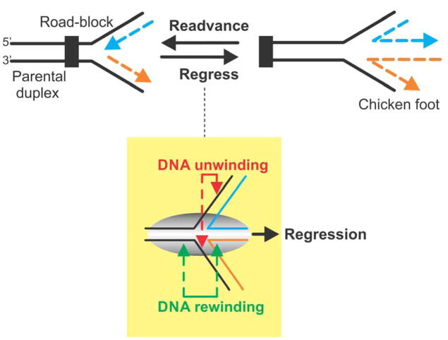
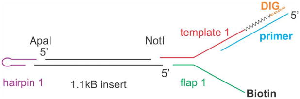
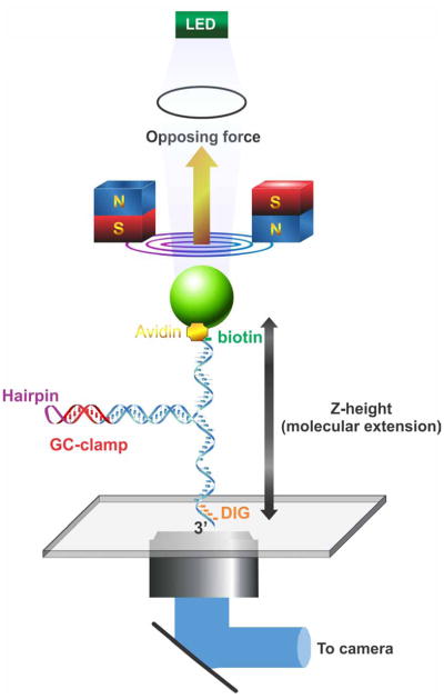
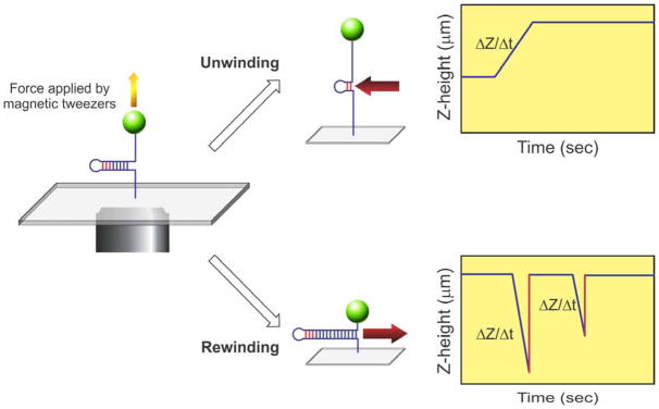
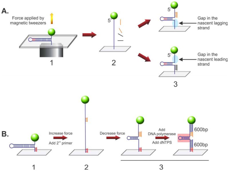
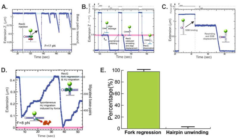
During DNA replication, forks often stall and require restart. One mechanism for restart requires that the fork be moved in a direction opposite to that of replication. This reaction is known as fork regression. For this reaction to occur, the enzyme must couple unwinding of the nascent heteroduplex fork arms to the rewinding of nascent strands ahead of itself and to the parental duplex in its wake. As the arms of the fork are complementary, this reaction is isoenergetic making it challenging to study. To overcome this, a novel adaptation of magnetic tweezers was developed by the Croquette group. Here, a 1200bp hairpin was attached at opposite ends to a flow cell surface and a magnetic bead. By manipulating the bead with the magnets, force can be applied to unwind the hairpin or alternatively, released to allow the hairpin to rewind. This adaptation was used to study fork regression by RecG. The results show that this is an efficient regression enzyme, able to work against a large opposing force. Critically, it couples DNA unwinding to duplex rewinding and in the process, can displace bound proteins from fork arms.

Keywords: DNA helicase; Fork regression; Hairpin substrate; Holliday junction; Magnetic tweezers; RecG; Replication fork; SSB.


Figures

Figure 1
Figure 1. Fork regression requires coupling of DNA unwinding to duplex rewinding
(A). A stalled replication fork is shown impeded and the nascent leading (blue) and lagging (orange) strands indicated. Fork regression results in rightward movement of the fork, away from the site of the replication road-block, concomitant with the extrusion of a duplex region resulting from the annealing of the nascent leading and lagging strands. The resulting DNA structure has been termed the “chicken foot” and is structurally equivalent to a Holliday junction. Fork readvancement occurs after regression and repair has taken place and results in leftward movement of the fork. Once complete, replisome reloading ensues. (B). Efficient regression requires the combined actions of nascent heteroduplex arm unwinding with DNA rewinding that occurs ahead of the advancing enzyme as well as in its wake. This results in reforming of the parental duplex and extrusion of a heteroduplex toe. The resulting three “toed” structure is known as the chicken foot intermediate.
Figure 2
Figure 2. DNA hairpin substrate construction
A schematic of the substrate is shown with each of the oligonucleotides shown. Sequences of each are provided in section 2.2. The purified insert from plasmid pNo-GTT is coloured black (center). The hairpin oligonucleotide (purple) is ligated to the 3′-overhang created by ApaI restriction enzyme cleavage. The annealed complex of flap 1 (green) and template 1 (red) is ligated to the 5′-overhang created by NotI restriction enzyme cleavage. Next, the primer (blue) is annealed to the 3′-end of template 1. Thereafter, T4 DNA polymerase and dNTPS including digoxgenin-labeled dUTP are added, extending the primer (zig-zag line) adding 6 DIG bases (orange) at the extreme 3′end of template 1.
Figure 3
Figure 3. Magnetic tweezers manipulate a novel fork substrate
A schematic of the 1,200 bp hairpin substrate under the careful control of magnetic tweezers is shown. The DNA molecule is held in place by site specific attachment to two surfaces: antibody-digoxygenin at the 3′-end (flow cell surface) and biotin-streptavidin at the 5′-end (bead surface). Tension in the DNA molecule and consequently its height (Z-extension) is carefully controlled by magnets of the tweezers, with opposing force directed away from the surface as indicated by the yellow arrow. Simultaneously, the bead is illuminated by an LED light source and the image captured by a CCD camera. Software is then used to calculate bead position (in nm) and to convert position data in DNA length (bp).
Figure 4
Figure 4. Key fork rescue reactions can be studied using a hairpin substrate
The hairpin substrate is held under tension by the magnetic tweezers (left panel). Introduction of enzyme and an energy source (ATP) can produce one of two reactions. If the enzyme catalyzes fork readvancement, it will preferentially unwind the duplex region of the substrate and the length of the DNA will increase as shown in the graph (top right). Thus the change in Z that occurs as a function of time can be attributed to the translocating enzyme. In contrast, if the enzyme catalyzes fork regression, motion will occur in the opposite direction (red arrow), and the two single stranded arms will be rewound (bottom right). Consequently, the net length of the DNA molecule will decrease as shown in the graph. In these reactions, duplex rewinding occurs and is terminated when the enzyme stalls permanently on the DNA or dissociates. This results in a virtually instantaneous increase in Z-height resulting from the opposing force applied by the magnetic tweezers (indicated by the red lines). The slope of the lines (ΔZ/Δt) is used to determine reaction rate and the length of the unwinding (or rewinding period) corresponds to processivity of the enzyme.
Figure 5
Figure 5. Stalled replication fork substrates can be constructed in situ
Two types of DNA substrates relevant to fork rescue can be constructed within the confines of the flow cell. (A) Construction of forks with gaps in either the leading or lagging strands. Here, the 1,200 bp hairpin is fully stretched by the application of force form the magnetic tweezers. Then, oligonucleotides complementary to the 5′- or 3′-proximal regions are introduced in separate reactions and allowed to bind. Once the opposing force is decreased, a partial hairpin is extruded and as the oligonucleotide remains annealed to reveal a fork with a gap on the opposite side, either the lagging (top) or leading strand (bottom). (B) Construction of a fork with duplex arms. As before the starting point is the 1,200 bp hairpin except now, a short DNA primer is annealed to 3′end of the substrate. Then, force is applied to unwind the hairpin and a second primer is introduced and allowed to bind. When the force is reduced, the DNA length decreases, a partial hairpin is extruded (~600bp in length) with primers bound to the opposing arms as indicated. When DNA polymerase is added, the tailed duplex regions are extended from each primer, producing a fork with 600 bp duplex arms. Pink box, parental duplex DNA region of the fork. The graph indicates the corresponding changes in Z-height as the hairpin is sequentially pulled apart and allow to reform as substrate construction proceeds.
Figure 6
Figure 6. RecG catalyzes the requisite reactions needed to regress a stalled replication fork
The data presented were published in (18) and are reused here in modified form. (A). RecG efficiently rewinds ssDNA fork arms. The DNA substrate is the core 1,200bp hairpin (Fig. 2 3). Reactions are done while the substrate is held under a constant tension of 17pN. Following the introduction of RecG multiple, separate events are observed where the extension of the DNA molecule decreases as a function of time. These events correspond to single, rewinding events where RecG reanneals the complementary arms. While the reactions rates are similar, the processivity of each event varies. (B). RecG couples DNA unwinding to duplex rewinding. In these reactions, the hairpin substrate is pulled apart by the application of force between t=118 and 122sec. Then an oligonucleotide is introduced and allowed to bind. Once the force is lowered, a partial hairpin is extruded to reveal a fork with a gap in the nascent lagging strand (Z-height indicated by the pink region). Once RecG is introduced, binding occurs, with the length of the on rate dictated by the nature of the fork (ton). The reaction then ensues and RecG unwinds the oligonucleotide resulting in its displacement, concomitant with rewinding of the ssDNA arms, resulting in extrusion of the hairpin. The hairpin can be repeatedly mechanically unfolded by the magnetic tweezers, allowing the displaced oligonucleotide to rebind and the reaction repeated. The length of ton was used to demonstrate substrate discrimination by RecG. For substrates with gaps in the leading strand ton= 15±1sec, whereas for forks with gaps in the lagging strand, ton= 1.8±0.1sec. (C). During rewinding, RecG displaces proteins bound to fork arms. Here, the hairpin was mechanically unfolded to the GC clamp and SSB (grey ovals) allowed to bind. As the protein wraps the ssDNA the net length of the molecule decreases. Once RecG is added, the extension decreased rapidly indicating fork arm rewinding, concomitant with SSB displacement. (D). RecG regresses a stalled fork resulting in formation of a Holliday junction. In these assays, the fork substrate was constructed using the scheme shown in Figure 4B. The resulting substrate is held in place by the magnetic tweezers using an opposing force of 8pN. Once RecG is introduced, extension rapidly decreases terminating at 0.08μm as RecG dissociates. As the now four arms of the resulting Holliday junction are equivalent, spontaneous junction migration occurs. When RecG rebinds, the reaction is repeated, this time at a slightly reduced rate (E). RecG catalyzes fork regression and not fork readvancement. A critical feature of the action of RecG at a fork is the direction in which it catalyzes fork movement. This was tested using the gapped substrates constructed in Figure 4A.

Similar articles

Cited by

References

    1. Kogoma T. Stable DNA replication: interplay between DNA replication, homologous recombination, and transcription. Microbiology & Molecular Biology Reviews. 1997;61(2):212–38. - PMC - PubMed
    1. Kuzminov A. Recombinational repair of DNA damage in Escherichia coli and bacteriophage lambda. Microbiol Mol Biol Rev. 1999;63(4):751–813. table of content. - PMC - PubMed
    1. Kowalczykowski SC. Initiation of genetic recombination and recombination-dependent replication. Trends in Biochemical Sciences. 2000;25(4):156–65. - PubMed
    1. Seigneur M, Bidnenko V, Ehrlich S, Michel B. RuvAB acts at arrested replication forks. Cell. 1998;95(3):419–30. - PubMed
    1. Cox MM, Goodman MF, Kreuzer KN, Sherratt DJ, Sandler SJ, Marians KJ. The importance of repairing stalled replication forks. Nature. 2000;404(6773):37–41. - PubMed

MeSH terms

LinkOut - more resources