

Severe muscle trauma triggers heightened and prolonged local musculoskeletal inflammation and impairs adjacent tibia fracture healing
- PMID: 27282456
- PMCID: PMC5114355
Severe muscle trauma triggers heightened and prolonged local musculoskeletal inflammation and impairs adjacent tibia fracture healing
Abstract
Objectives: Complicated fracture healing is often associated with the severity of surrounding muscle tissue trauma. Since inflammation is a primary determinant of musculoskeletal health and regeneration, it is plausible that delayed healing and non-unions are partly caused by compounding local inflammation in response to concomitant muscle trauma.
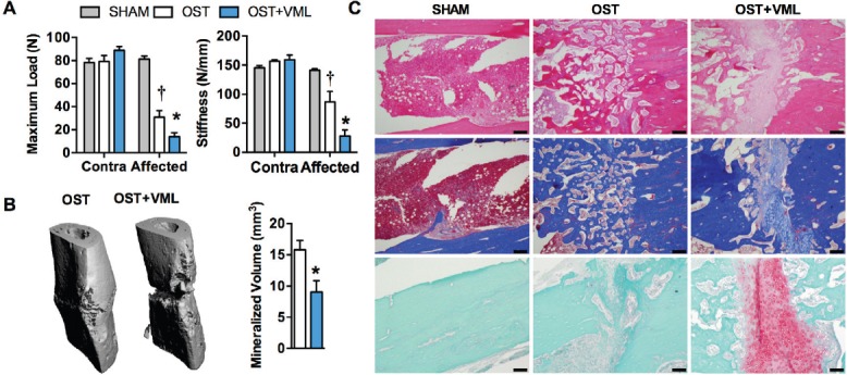
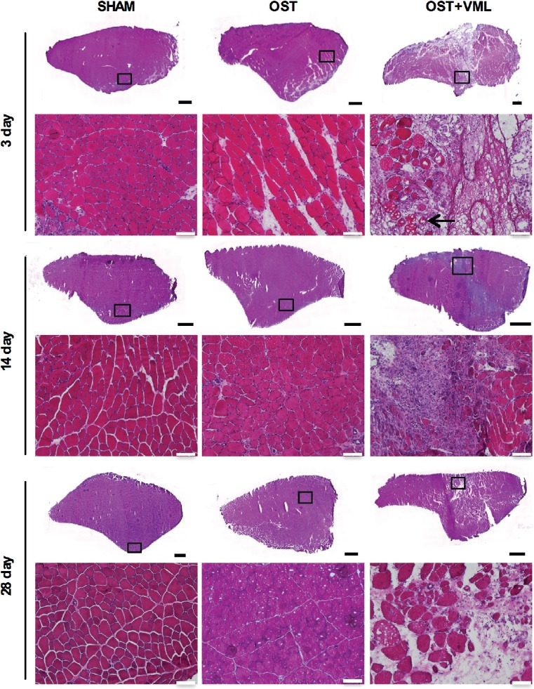
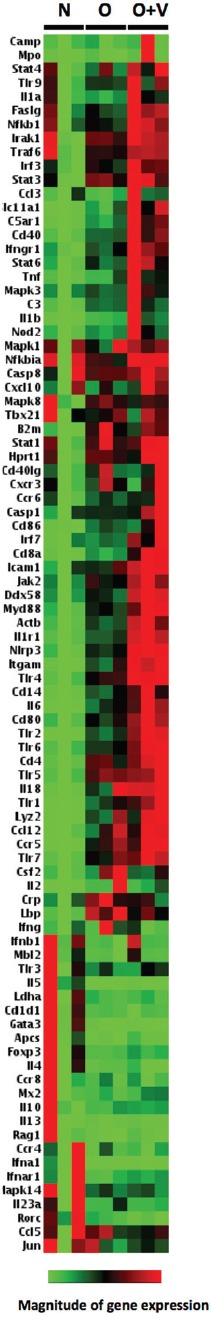
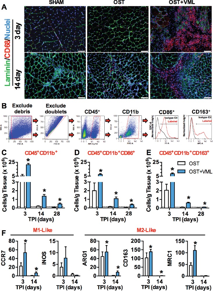
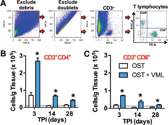
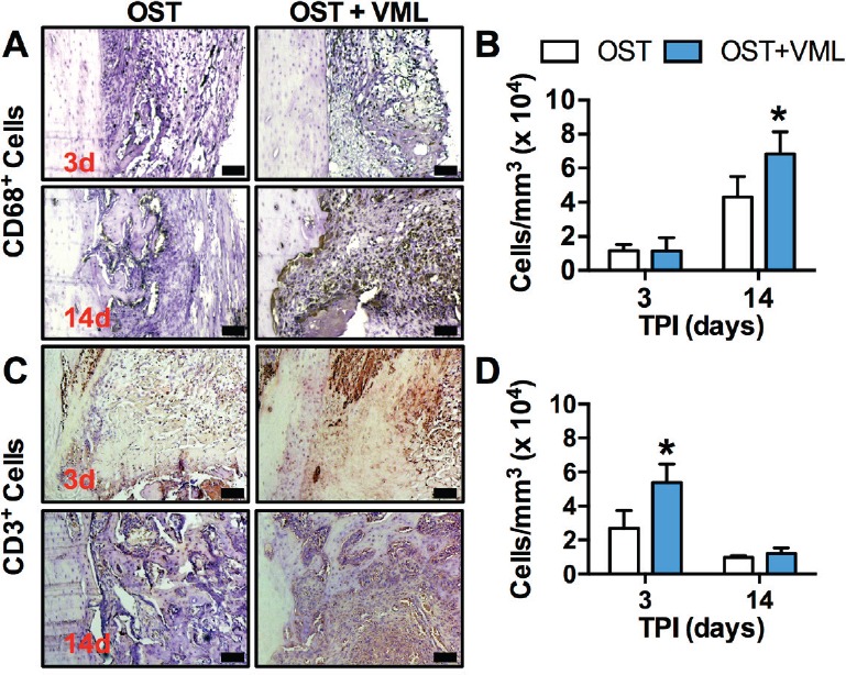
Methods and results: To investigate this possibility, a Lewis rat open fracture model [tibia osteotomy with adjacent tibialis anterior (TA) muscle volumetric muscle loss (VML) injury] was interrogated. We observed that VML injury impaired tibia healing, as indicated by diminished mechanical strength and decreased mineralized bone within the fracture callus, as well as continued presence of cartilage instead of woven bone 28 days post-injury. The VML injured muscle presented innate and adaptive immune responses that were atypical of canonical muscle injury healing. Additionally, the VML injury resulted in a perturbation of the inflammatory phase of fracture healing, as indicated by elevations of CD3(+) lymphocytes and CD68+ macrophages in the fracture callus at 3 and 14d post-injury, respectively.
Conclusions: These data indicate that heightened and sustained innate and adaptive immune responses to traumatized muscle are associated with impaired fracture healing and may be targeted for the prevention of delayed and non-union following musculoskeletal trauma.
Conflict of interest statement
The authors have no conflict of interest. The opinions or assertions contained herein are the private views of the authors and are not to be construed as official or as reflecting the views of the Department of the Army or the Department of Defense.
Figures
Similar articles
-
Impairment of early fracture healing by skeletal muscle trauma is restored by FK506.BMC Musculoskelet Disord. 2017 Jun 12;18(1):253. doi: 10.1186/s12891-017-1617-y. BMC Musculoskelet Disord. 2017. PMID: 28606129 Free PMC article.
-
Autologous minced muscle grafts improve endogenous fracture healing and muscle strength after musculoskeletal trauma.Physiol Rep. 2017 Jul;5(14):e13362. doi: 10.14814/phy2.13362. Physiol Rep. 2017. PMID: 28747511 Free PMC article.
-
Decellularized extracellular matrix repair of volumetric muscle loss injury impairs adjacent bone healing in a rat model of complex musculoskeletal trauma.J Trauma Acute Care Surg. 2016 Nov;81(5 Suppl 2 Proceedings of the 2015 Military Health System Research Symposium):S184-S190. doi: 10.1097/TA.0000000000001212. J Trauma Acute Care Surg. 2016. PMID: 27533905
-
Time Course of Immune Response and Immunomodulation During Normal and Delayed Healing of Musculoskeletal Wounds.Front Immunol. 2020 Jun 4;11:1056. doi: 10.3389/fimmu.2020.01056. eCollection 2020. Front Immunol. 2020. PMID: 32582170 Free PMC article. Review.
-
The use of gentamicin-coated nails in complex open tibia fracture and revision cases: A retrospective analysis of a single centre case series and review of the literature.Injury. 2015 Dec;46(12):2433-7. doi: 10.1016/j.injury.2015.09.028. Epub 2015 Oct 8. Injury. 2015. PMID: 26477343 Review.
Cited by
-
The Role of Innate and Adaptive Immune Cells in Skeletal Muscle Regeneration.Int J Mol Sci. 2021 Mar 23;22(6):3265. doi: 10.3390/ijms22063265. Int J Mol Sci. 2021. PMID: 33806895 Free PMC article. Review.
-
Resistance wheel running improves contractile strength, but not metabolic capacity, in a murine model of volumetric muscle loss injury.Exp Physiol. 2023 Oct;108(10):1282-1294. doi: 10.1113/EP091284. Epub 2023 Aug 10. Exp Physiol. 2023. PMID: 37526646 Free PMC article.
-
The local and systemic effects of immune function on fracture healing.OTA Int. 2024 Mar 11;7(2 Suppl):e328. doi: 10.1097/OI9.0000000000000328. eCollection 2024 Mar. OTA Int. 2024. PMID: 38487403 Free PMC article. Review.
-
Response of terminal Schwann cells following volumetric muscle loss injury.Exp Neurol. 2023 Jul;365:114431. doi: 10.1016/j.expneurol.2023.114431. Epub 2023 May 2. Exp Neurol. 2023. PMID: 37142114 Free PMC article.
-
Effects of experimental cervical spinal cord injury on peripheral adaptive immunity.PLoS One. 2020 Oct 30;15(10):e0241285. doi: 10.1371/journal.pone.0241285. eCollection 2020. PLoS One. 2020. PMID: 33125407 Free PMC article.
References
-
- MacKenzie EJ, Bosse MJ. Factors influencing outcome following limb-threatening lower limb trauma:lessons learned from the Lower Extremity Assessment Project (LEAP) J Am Acad Orthop Surg. 2006;14:S205–10. - PubMed
-
- Doukas WC, Hayda RA, Frisch HM, et al. The Military Extremity Trauma Amputation/Limb Salvage (METALS) study:outcomes of amputation versus limb salvage following major lower-extremity trauma. J Bone Joint Surg Am. 2013;95:138–45. - PubMed
-
- Corona BT, Garg K, Ward CL, McDaniel JS, Walters TJ, Rathbone CR. Autologous minced muscle grafts:a tissue engineering therapy for the volumetric loss of skeletal muscle. Am J Physiol Cell Physiol. 2013;305:C761–75. - PubMed
-
- Garg K, Corona BT, Walters TJ. Losartan administration reduces fibrosis but hinders functional recovery after volumetric muscle loss injury. J Appl Physiol (1985) 2014;117:1120–31. - PubMed
-
- Garg K, Ward CL, Hurtgen BJ, et al. Volumetric muscle loss:persistent functional deficits beyond frank loss of tissue. J Orthop Res. 2015;33:40–6. - PubMed