. 2016 Jun 10:6:27512.
doi: 10.1038/srep27512.

Mechanisms of PDGF siRNA-mediated inhibition of bone cancer pain in the spinal cord

Affiliations

Mechanisms of PDGF siRNA-mediated inhibition of bone cancer pain in the spinal cord

Yang Xu et al. Sci Rep. .

Abstract

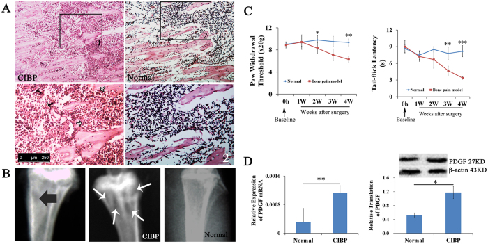
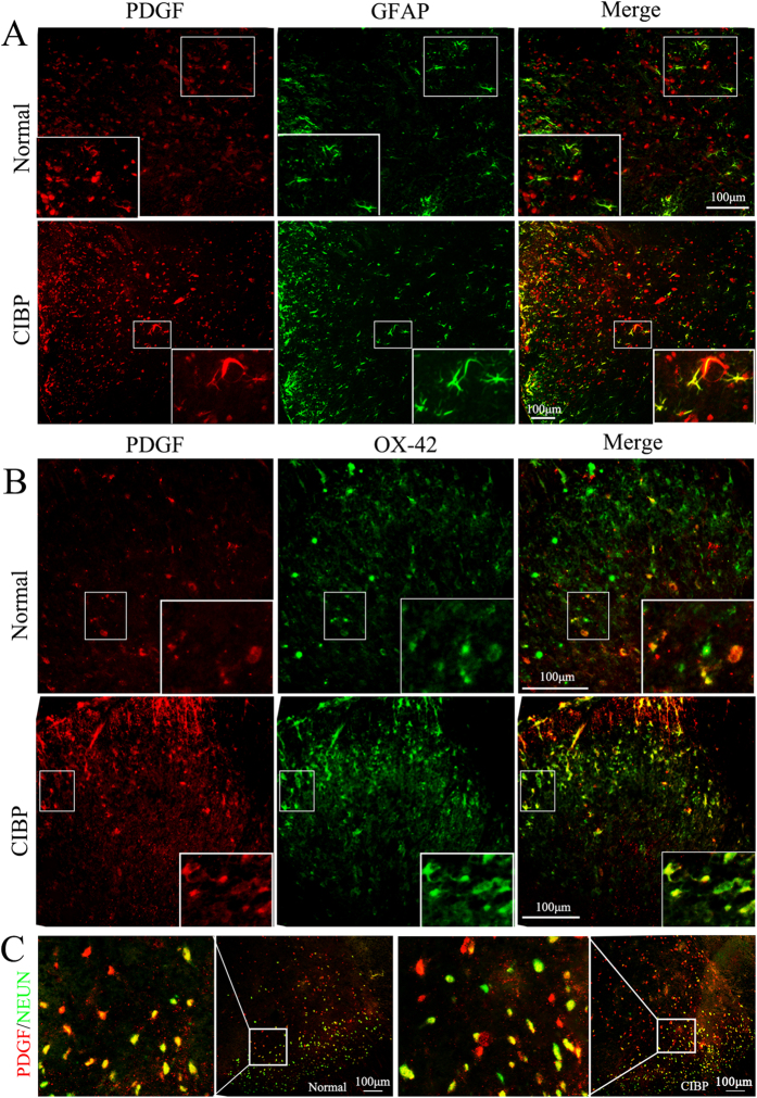
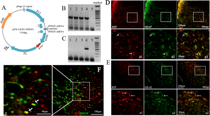
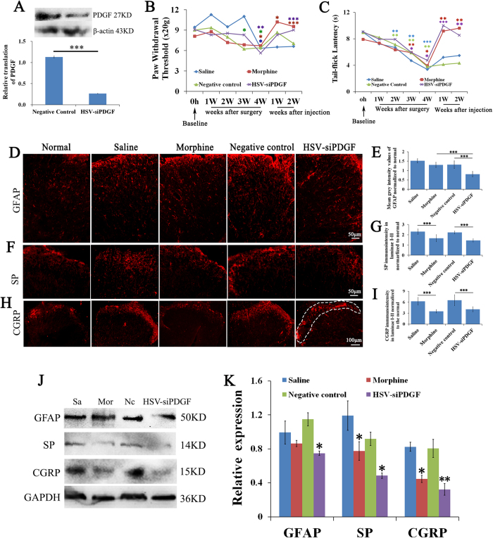
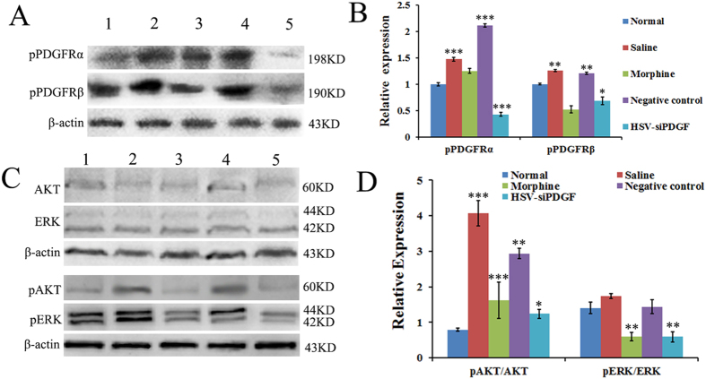
Patients with tumors that metastasize to bone frequently suffer from debilitating pain, and effective therapies for treating bone cancer are lacking. This study employed a novel strategy in which herpes simplex virus (HSV) carrying a small interfering RNA (siRNA) targeting platelet-derived growth factor (PDGF) was used to alleviate bone cancer pain. HSV carrying PDGF siRNA was established and intrathecally injected into the cavum subarachnoidale of animals suffering from bone cancer pain and animals in the negative group. Sensory function was assessed by measuring thermal and mechanical hyperalgesia. The mechanism by which PDGF regulates pain was also investigated by comparing the differential expression of pPDGFRα/β and phosphorylated ERK and AKT. Thermal and mechanical hyperalgesia developed in the rats with bone cancer pain, and these effects were accompanied by bone destruction in the tibia. Intrathecal injection of PDGF siRNA and morphine reversed thermal and mechanical hyperalgesia in rats with bone cancer pain. In addition, we observed attenuated astrocyte hypertrophy, down-regulated pPDGFRα/β levels, reduced levels of the neurochemical SP, a reduction in CGRP fibers and changes in pERK/ERK and pAKT/AKT ratios. These results demonstrate that PDGF siRNA can effectively treat pain induced by bone cancer by blocking the AKT-ERK signaling pathway.


Figures

Figure 1
Figure 1. Bone cancer-induced bone destruction, thermal and mechanical hyperalgesia, and increased PDGF expression.
(A) Hematoxylin and eosin (H&E) staining of the tibia revealed that bone marrow spaces were infiltrated with malignant tumor cells (black arrows) and osteoclasts (white arrows) on day 28 after the injection of tumor cells. The images in the lower panel are magnified images of the boxes in the images in the upper panel. (B) High-resolution radiographs of rat hind limbs after the bilateral injection of tumor cells into the rostral part of the tibia. The left image indicates the site of tumor cell injection (black arrow), and the middle image indicates areas of bone destruction (white arrows). (C) Paw withdrawal threshold and tail-flick latency were evaluated over the indicated time course. (D) Animals with bone cancer pain exhibited elevated expression levels of PDGF as measured by qPCR and western blot. Data are presented as the mean ± SEM. *P < 0.05, **P < 0.01, ***P < 0.001.
Figure 2
Figure 2. Enhanced expression of PDGF in neurons, astrocytes and microglia cells in the spinal cords of rats with bone cancer pain compared with normal rats.
(A,B,C) Double immunofluorescence labeling in the CIBP and normal groups demonstrated that PDGF (red) localized to neurons, astrocytes and microglia cells in the dorsal horn of the spinal cord. The inset of each image is a magnified view of the indicated region. NEUN: neuron marker (green); GFAP: astrocyte marker (green); OX-42: microglia maker (green).
Figure 3
Figure 3. The PDGF siRNA replication-incompetent HSV-1 vector.
(A) Schematic representation of the HSV-1-based gene transfer vector. The construct was created by deleting the 3 immediate-early genes (ICP4, ICP27 and ICP34.5) of wild-type HSV-1. The vector also contained the human cytomegalovirus (CMV) promoter, WPRE and a polyA (pA) region. (B) The presence of the desired elements in the vector, denoted pNX01-U6H1-rPDGFi, was confirmed by RT-PCR analysis. Markers (100 bp DNA ladder, from top to bottom): 1,500, 1,000, 900, 800, 700, 600, 500, 400, 300, 200 and 100 bp fragments. (C) The production of NX01-U6H1-rPDGFi was confirmed by RT-PCR analysis. (D–F) Cells infected with the HSV virus were analyzed by double immunofluorescence labeling 7 days after the intrathecal injections. Panels d1–d3, e1–e3 and f are magnified views of the boxes in D–F, respectively, and demonstrate the colocalization for RFP (red) with GFAP (green), OX-42 (green) and NEUN (green). RFP is a marker protein in the HSV vector.
Figure 4
Figure 4. PDGF siRNA down regulates PDGF protein levels, attenuates tumor-evoked mechanical and thermal hyperalgesia, reverses bone cancer pain-evoked astrocyte hypertrophy and reduces the expression of substance P (SP) and CGRP in laminae I-II of L4–6 in the dorsal horn of the spinal cord.
(A) Treatment with PDGF siRNA down-regulated the transcription of PDGF. (B) Paw withdrawal thresholds for each treatment groups were evaluated throughout the study. (C) The tail-flick latencies of each treatment group were evaluated throughout the study. (D,F,H) Images of the dorsal horn showing the distribution of the astrocyte markers GFAP, SP and CGRP in transverse sections at spinal cord segments L4–L6 in animals that received saline, morphine, negative control siRNA and PDGF siRNA. (E) Quantification of GFAP immunofluorescence demonstrated that treatment with PDGF siRNA reduced GFAP-immunoreactivity in tumor-injected rats. (G,I) Quantification of SP and CGRP in laminae I-II revealed a reduction following treatment with PDGF siRNA or morphine. DAPI: nuclear marker (blue). The white dashed area shows laminae I-II of the spinal cord. (J, K) The protein levels of GFAP, SP and CGRP were consistent with the results of immunofluorescence. Symbols with different colors represent different groups. Data are presented as the mean ± SEM. *P < 0.05, **P < 0.01, ***P < 0.001.
Figure 5
Figure 5. Bone cancer pain increases phosphorylation of PDGFRs, ERK and AKT in the dorsal horn of spinal cord L4–6 segments, and PDGF siRNA reverses this effect.
(A–D) Western blot was performed to evaluate the levels of phosphorylated PDGFRs (A,B), AKT and extracellular regulated kinase (ERK)1/2 (C,D) in protein extracts from the L4–6 segment of the spinal cord in each group. The level of pPDGFRs decreased in the PDGF siRNA-treated group (A,B). Treatment with PDGF siRNA and morphine significantly reduced the pERK/ERK and pAKT/AKT ratios (C,D). β-actin, a house-keeping gene was used as an internal control. Lane 1: Normal; Lane 2: Treatment with saline; Lane 3: Treatment with morphine; Lane 4: Treatment with negative control siRNA; Lane 5: Treatment with PDGF siRNA. Data are represented as the mean ± SEM. *P < 0.05, **P < 0.01, ***P < 0.001.

Similar articles

Cited by

References

    1. Coleman R. E. Skeletal complications of malignancy. Cancer. 80, 1588–1594 (2004). - PubMed
    1. Lipton A. et al. Pamidronate prevents skeletal complications and is effective palliative treatment with breast carcinoma and osteolytic bone metastases: long-term follow-up of two randomized, placebo-controlled trails. Cancer. 88, 1082–1090 (2000). - PubMed
    1. Bernard W. S. & Christopher P. W. The global and regional burden of cancer in World cancer report 2014. (eds Devid F. et al.) 26–27 (International Agency for Research on Cancer, 2014).
    1. Coleman R. E. Clinical features of metastatic bone disease and risk of skeletal morbidity. Clin Cancer Res. 12, 6243s–6249s (2006). - PubMed
    1. Galasko C. S. B. [The anatomy and pathways of skeletal metastases] Bone Metastases [Weiss L. & Gilbert A. (ed.)] [49–63] (Boston, GK Hall, 1981).

Publication types

MeSH terms

Substances