. 2016 Jul 19;12(8):2417-26.
doi: 10.1039/c6mb00304d.

Histidine switch controlling pH-dependent protein folding and DNA binding in a transcription factor at the core of synthetic network devices

Affiliations

Histidine switch controlling pH-dependent protein folding and DNA binding in a transcription factor at the core of synthetic network devices

D K Deochand et al. Mol Biosyst. .

Abstract

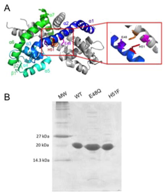
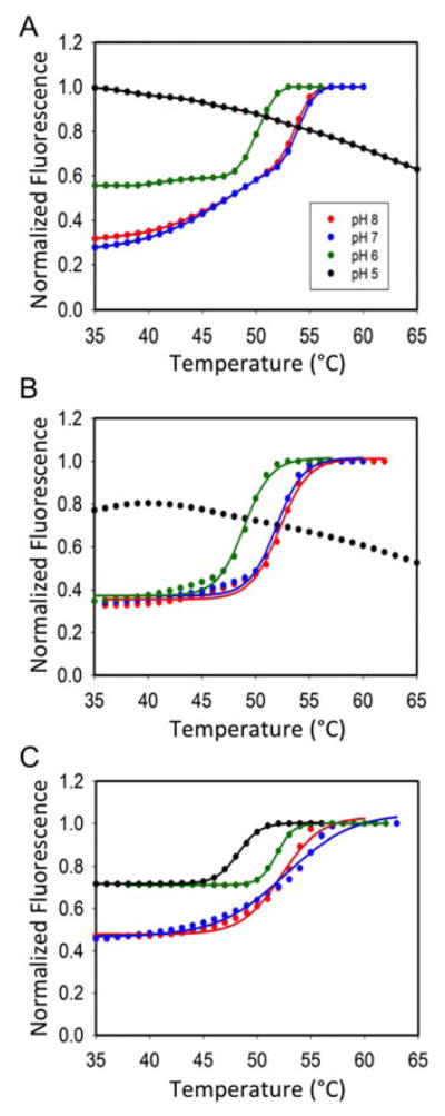
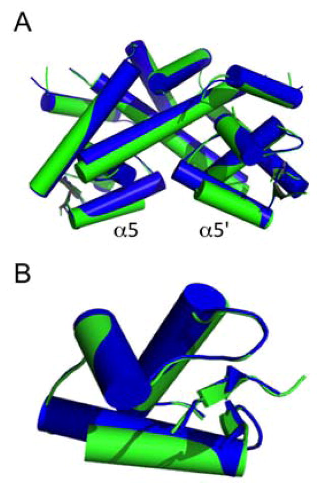
Therapeutic strategies have been reported that depend on synthetic network devices in which a urate-sensing transcriptional regulator detects pathological levels of urate and triggers production or release of urate oxidase. The transcription factor involved, HucR, is a member of the multiple antibiotic resistance (MarR) protein family. We show that protonation of stacked histidine residues at the pivot point of long helices that form the scaffold of the dimer interface leads to reversible formation of a molten globule state and significantly attenuated DNA binding at physiological temperatures. We also show that binding of urate to symmetrical sites in each protein lobe is communicated via the dimer interface. This is the first demonstration of regulation of a MarR family transcription factor by pH-dependent interconversion between a molten globule and a compact folded state. Our data further suggest that HucR may be utilized in synthetic devices that depend on detection of pH changes.


Figures

Figure 1
Figure 1
HucR variants. A. Stacked H51/H51′ at the dimer interface of HucR-WT. Cartoon representation of HucR-WT with one monomer colored blue to green (N- to C-terminus) and the other monomer in gray. The close-up view shows H51/H51′ in red and orange and neighboring E48/E48′ in magenta. B. Purified HucR-WT, HucR-E48Q, and HucR-H51F in 15% SDS-PAGE gel. Molecular weight (MW) markers are shown at the left.
Figure 2
Figure 2
Thermal stability determined by fluorometry. Normalized fluorescence of SYPRO Orange bound to hydrophobic patches of unfolded protein/molten globule states as a function of temperature at pH 8.0 (red), 7.0 (blue), 6.0 (green), and 5.0 (black). A. HucR-WT. B. HucR-E48Q. C. HucR-H51F.
Figure 3
Figure 3
CD spectral analysis of HucR variants. A. CD spectra of HucR-WT (black), HucR-E48Q (red), and HucR-H51F (green) at pH 8.0. B. CD spectra of HucR-WT (black), HucR-E48Q (red), and HucR-H51F (green) at pH 5.0. C–D. Thermal stability of HucR variants at pH 5.0. Ellipticity was recorded at 218–224 nm in CD2 buffer (50 mM sodium phosphate pH 7.0 or 50 mM acetate pH 5.0, both with 100 mM NaCl). C. HucR-WT (black) and HucR-E48Q (red). D. HucR-H51F.
Figure 4
Figure 4
DNA binding affinity at pH 8.0. A–C. Electrophoretic mobility shift assay of HucR-WT, HucR-E48Q, and HucR-H51F binding to 0.1 nM hucO (77-bp) at room temperature. The sequence of the HucR binding site is shown in Fig. S2 (red). Complex (C) and free DNA (D) are indicated at the left. D. Binding isotherms for HucR-WT (continuous line) and HucR-E48Q (broken line). E. Binding isotherm for HucR-H51F. Error bars represent standard deviation from three independent experiments.
Figure 5
Figure 5
Thermal denaturation of HucR variants incubated with urate at pH 8.0. A–C. Thermal denaturation measured by normalized SYPRO Orange fluorescence. A. HucR-WT. B. HucR-E48Q. C. HucR-H51F. D–E. Thermal stability of HucR-WT measured by CD spectroscopy at 218–224 nm. D. HucR-WT supplemented with 0.1 M NaOH (the solvent for urate). E. HucR-WT with 10 μM urate (blue) or 50 μM urate (green).
Figure 6
Figure 6
Structural comparison between HucR-WT and HucR-E48Q. A. Superimposition of HucR-WT (blue) and HucR-E48Q (green). The long, central α2 helices form the scaffold of the dimer interface and residues H51 and H51′ are located where α2 helices intersect, as shown in Fig. 1. B. The DNA binding domain showing significant conformational change in the DNA binding helix (α5).

Similar articles

Cited by

References

    1. Nandagopal N, Elowitz MB. Science. 2011;333:1244–1248. - PMC - PubMed
    1. Weber W, Fussenegger M. Nat Rev Genet. 2012;13:21–35. - PMC - PubMed
    1. Ye H, Fussenegger M. FEBS Lett. 2014;588:2537–2544. - PubMed
    1. Kemmer C, Gitzinger M, Daoud-El Baba M, Djonov V, Stelling J, Fussenegger M. Nat Biotechnol. 2010;28:355–360. - PubMed
    1. Geraths C, Daoud-El Baba M, Charpin-El Hamri G, Weber W. J Control Release. 2013;171:57–62. - PubMed

Publication types

LinkOut - more resources