. 2016 Nov;37(11):1983-1991.
doi: 10.3174/ajnr.A4851. Epub 2016 Jun 9.

Bidirectional Changes in Anisotropy Are Associated with Outcomes in Mild Traumatic Brain Injury

Affiliations

Bidirectional Changes in Anisotropy Are Associated with Outcomes in Mild Traumatic Brain Injury

S B Strauss et al. AJNR Am J Neuroradiol. 2016 Nov.

Abstract

Background and purpose: Mild traumatic brain injury results in a heterogeneous constellation of deficits and symptoms that persist in a subset of patients. This prospective longitudinal study identifies early diffusion tensor imaging biomarkers of mild traumatic brain injury that significantly relate to outcomes at 1 year following injury.

Materials and methods: DTI was performed on 39 subjects with mild traumatic brain injury within 16 days of injury and 40 controls; 26 subjects with mild traumatic brain injury returned for follow-up at 1 year. We identified subject-specific regions of abnormally high and low fractional anisotropy and calculated mean fractional anisotropy, axial diffusivity, radial diffusivity, and mean diffusivity across all white matter voxels brain-wide and each of several white matter regions. Assessment of cognitive performance and symptom burden was performed at 1 year.

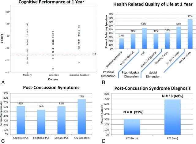
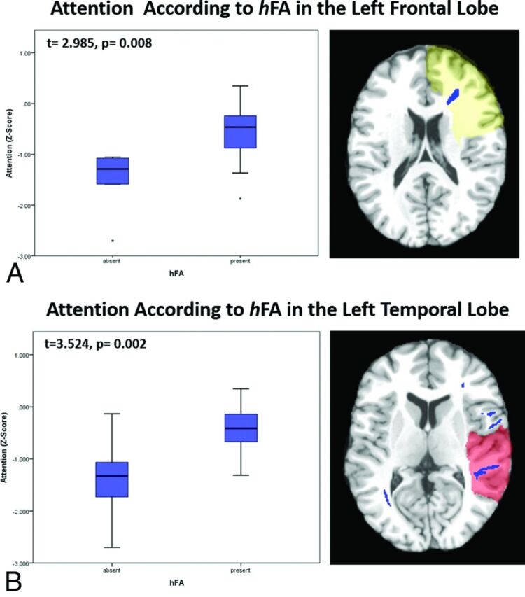
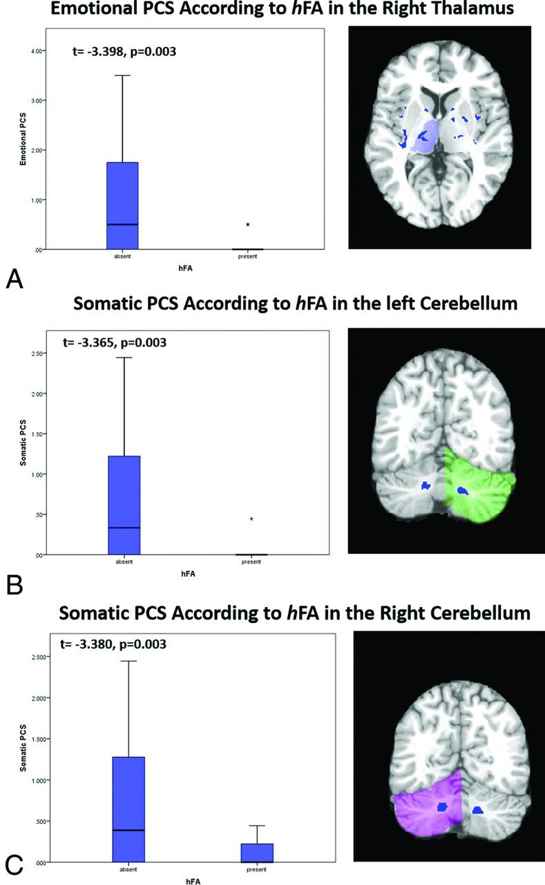
Results: Significant associations of brain-wide DTI measures and outcomes included the following: mean radial diffusivity and mean diffusivity with memory; and mean fractional anisotropy, radial diffusivity, and mean diffusivity with health-related quality of life. Significant differences in outcomes were found between subjects with and without abnormally high fractional anisotropy for the following white matter regions and outcome measures: left frontal lobe and left temporal lobe with attention at 1 year, left and right cerebelli with somatic postconcussion symptoms at 1 year, and right thalamus with emotional postconcussion symptoms at 1 year.

Conclusions: Individualized assessment of DTI abnormalities significantly relates to long-term outcomes in mild traumatic brain injury. Abnormally high fractional anisotropy is significantly associated with better outcomes and might represent an imaging correlate of postinjury compensatory processes.


Figures

Fig 1.
Fig 1.
Outcomes in mTBI. A, Cognitive outcomes. The red dotted line indicates impairment in the given domain, defined as a z score less than −1.5. B, Health-related quality of life (Sickness Impact Profile), grouped according to dimension. C, Postconcussion symptoms (Rivermead Post Concussion Symptoms Questionnaire). D, Postconcussion syndrome diagnosis. PAC indicates psychological autonomy and communication.
Fig 2.
Fig 2.
Associations between regional imaging measures and long-term cognitive outcomes. A, Subjects with high FA in the left frontal lobe perform significantly better on tasks of attention at 1 year postinjury than do those without it (t = 2.985, P = .008). B, Subjects with high FA in left temporal lobe perform significantly better on tasks of attention at 1 year postinjury than do those without it (t = 3.524, P = .002).
Fig 3.
Fig 3.
Associations between regional imaging measures and long-term functional outcomes. A, Subjects with hFA in the right thalamus have significantly fewer emotional postconcussion symptoms at 1 year than those without it (t = −3.398, P = .003). Of those with hFA in the right thalamus, only 2 subjects reported emotional postconcussion symptoms. The asterisk indicates an outlier. B, Subjects with hFA in the left cerebellar white matter have significantly fewer somatic postconcussion symptoms at 1 year than those without hFA in the left cerebellum (t = −3.365, P = .003). Of those with hFA in the left cerebellum, only 1 subject reported somatic postconcussion symptoms. The asterisk indicates an outlier. C, Subjects with hFA in the right cerebellar white matter have significantly fewer somatic postconcussion symptoms at 1 year than do those without high FA in the right cerebellum (t = −3.380, P = .003). Of those with hFA in the left cerebellum, only 2 reported somatic postconcussion symptoms.

References

    1. Ruff R. Two decades of advances in understanding of mild traumatic brain injury. J Head Trauma Rehabil 2005;20:5–18 10.1097/00001199-200511000-00009 - DOI - PubMed
    1. Bigler ED. Neuropsychological results and neuropathological findings at autopsy in a case of mild traumatic brain injury. J Int Neuropsychol Soc 2004;10:794–806 - PubMed
    1. Johnson VE, Stewart W, Smith DH. Axonal pathology in traumatic brain injury. Exp Neurol 2013;246:35–43 10.1016/j.expneurol.2012.01.013 - DOI - PMC - PubMed
    1. Mac Donald C, Dikranian K, Song S, et al. Detection of traumatic axonal injury with diffusion tensor imaging in a mouse model of traumatic brain injury. Exp Neurol 2007;205:116–31 10.1016/j.expneurol.2007.01.035 - DOI - PMC - PubMed
    1. Shenton M, Hamoda H, Schneiderman J, et al. A review of magnetic resonance imaging and diffusion tensor imaging findings in mild traumatic brain injury. Brain Imaging Behav 2012;6:137–92 10.1007/s11682-012-9156-5 - DOI - PMC - PubMed

LinkOut - more resources