. 2016 Jun 10:6:26351.
doi: 10.1038/srep26351.

Up-regulation of PKM2 promote malignancy and related to adverse prognostic risk factor in human gallbladder cancer

Affiliations

Up-regulation of PKM2 promote malignancy and related to adverse prognostic risk factor in human gallbladder cancer

Wei Lu et al. Sci Rep. .

Abstract

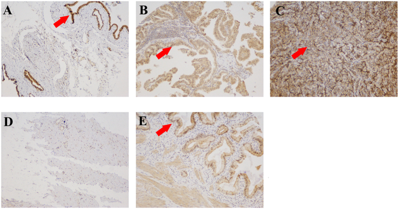
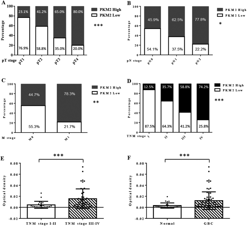
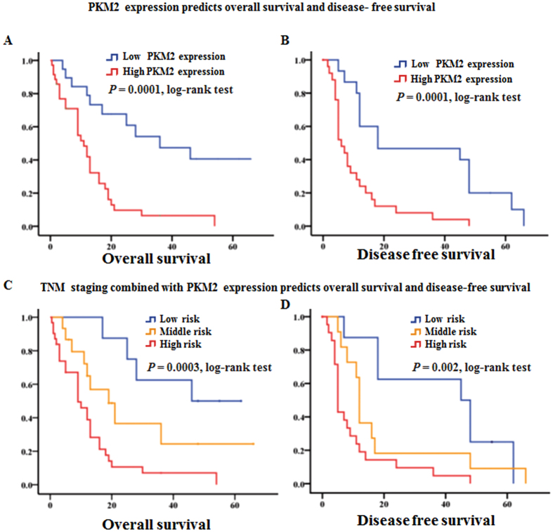
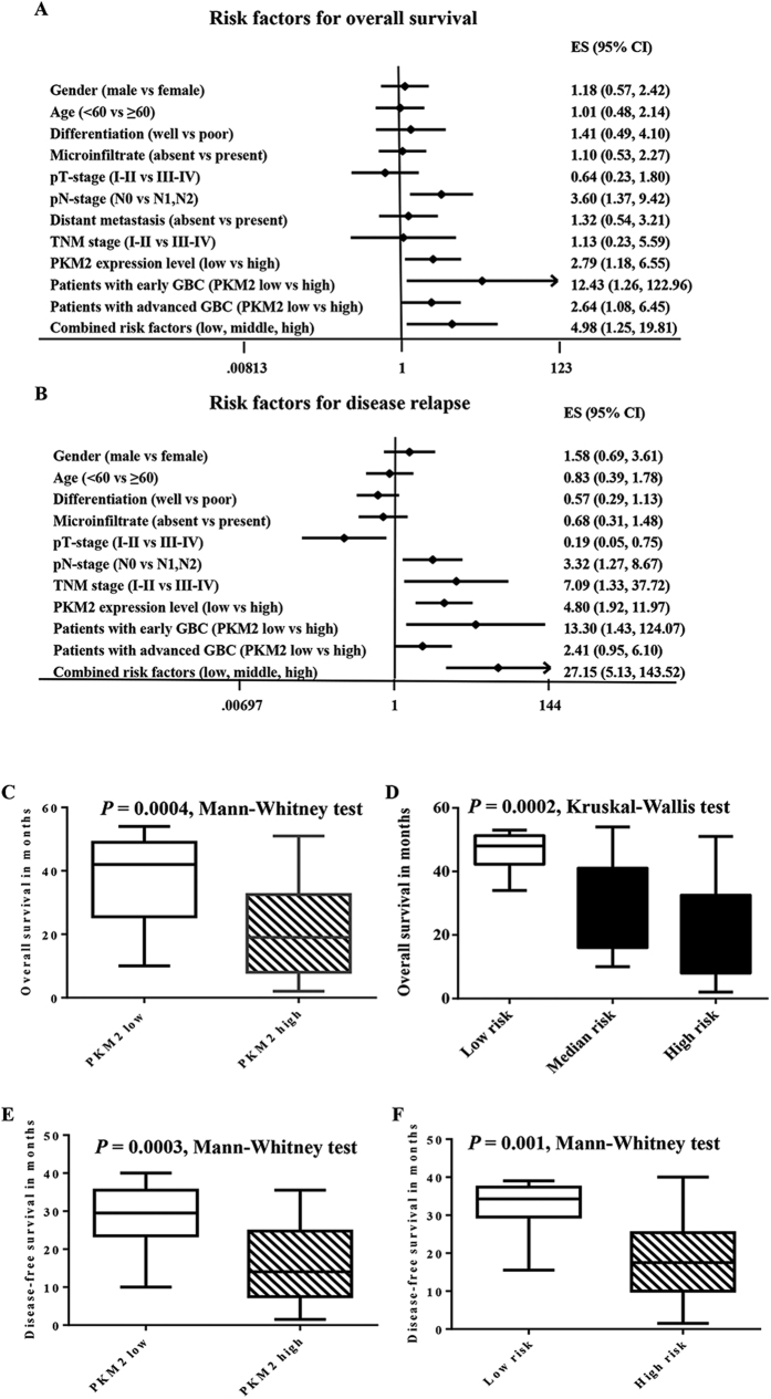
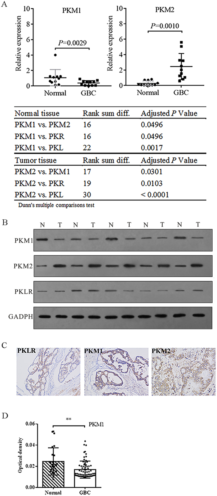
Recently, pyruvate kinase M2 (PKM2) has been implicated in the progression of certain cancers and might play pivotal roles in the formation of malignancy. However, the role of PKM2 in gallbladder cancer had not been well investigated. This study analyzed associations between PKM2 expression status with various clinical and pathologic parameters in a large cohort of gallbladder cancer (GBC) patients from a long term follow up results. The expression level of pyruvate kinase isotypes in GBC tissues and their adjacent normal gallbladder tissues were estimated by qRT-PCR and Western blot. PKM2 mRNA level were significantly high in gallbladder cancer tissues than in adjacent noncancerous tissues (P < 0.001). High expression of the PKM2 was detected in 55.71% paraffin-embedded GBC tissue. The high PKM2 expression was independently associated with poorer overall survival in patients with GBC (median survival 11.9 vs 30.1 months; hazard ratio 2.79; 95% CI = 1.18 to 6.55; P = 0.02). These findings indicated elevated expression of PKM2 is a prognostic factor for poor GBC clinical outcomes, implied involving of PKM2 in GBC progression.


Figures

Figure 1
Figure 1. PKM2 expression in sections of cancerous gallbladder tissue.
(A) Well-differentiated adenocarcinoma exhibited low PKM2 staining and was given a score of 2. (B) Moderately differentiated adenocarcinoma exhibited moderate PKM2 expression and was given a score of 3. (C) Poorly differentiated adenocarcinoma exhibited strong PKM2 expression and was given a score of 4. (D) Gallbladder tissue was used as the negative control as it exhibited negative staining for PKM2 and was given a score of 0. (E) Gallbladder tissue which were positive staining for PKM2 and was given a score of 1 (F) Western blot with an antiPKM2 antibody revealed positive expression of PKM2 in GBC-SD, NOZ and SGC-996 cells. The red arrow in (AC,E) point to the positive stained cell which were dyed brown. Original magnification: ×200.
Figure 2
Figure 2. The PKM2 expression level in gallbladder cancer tissues was significantly higher than in normal gallbladder tissues and was associated with clinical stage.
(A–D) The percentage of patients with low PKM2 expression decreased gradually as the disease progressed from pT stage 1 to 4 and pN stage 0 to 3 and when non-metastatic disease progressed to metastatic disease; percentages are also summarized according to TNM stages I to IV. (E) Comparison of the dot distribution of the optical density of PKM2 protein staining in gallbladder tumor tissues according to the TNM stage (TNM I–II are regarded as early stage, and III–IV are regarded as advanced stage; ***P = 0.0008, Mann-Whitney U test). (F) Comparison of the dot distribution of the optical density of PKM2 protein staining in gallbladder tumor tissues with normal gallbladder tissues ***P = 0.0007, Mann-Whitney U test).
Figure 3
Figure 3. PKM2 expression level and the combined risk factor score predict OS and DFS in GBC patients.
(A,B) Kaplan-Meier survival estimates revealed that high PKM2 immunoreactivity in gallbladder adenocarcinoma was significantly associated with worse overall survival and worse disease-free survival according to the dichotomy of the extent of PKM2 staining intensity. (C,D) Kaplan-Meier survival estimates showed that the trichotomy of the combined risk scores was significantly associated with corresponding overall survival prognosis. A high-risk score represents worse overall survival (OS) and disease-free survival (DFS), whereas a low-risk score represents relatively improved OS and DFS rates. However, a median-risk score predicts median OS but worse DFS.
Figure 4
Figure 4. Analyses of risk factors in gallbladder cancer patients.
(A) A multivariate Cox analysis of the clinical and pathological factors showed predictive value with respect to OS in gallbladder cancer patients. Generally, PKM2 and pN stage showed prognostic value for predicting OS. Specifically, PKM2 demonstrated prominent prognostic value in patients with early stage gallbladder cancer (i.e., TNM stages I and II), and the combination of the TNM stages and the PKM2 staining demonstrated better predictive value than either alone. (B) A multivariate Cox analysis of the clinical and pathological factors demonstrated predictive value with respect to DFS in gallbladder cancer patients. Generally, PKM2 and pN stage showed prognostic value for predicting DFS. Specifically, PKM2 exhibited better prognostic value in patients with early stage gallbladder cancer (i.e., TNM stages I and II). The combination of TNM stage and PKM2 staining showed better predictive value than either alone. (C–F) A rank-sum test compared PKM2 expression levels and the combined risk factors to predict OS and DFS.
Figure 5
Figure 5. The pyruvate isotypes expression in gallbladder cancer tissues and normal gallbladder tissues.
(A) Compare the expression of PKM1 and PKM2 mRNA in gallbladder cancer tissue with adjacent normal gallbladder tissues. (B) Compare the protein level of pyruvate kinase isotypes in 5 paired gallbladder cancer tissues and normal counterpart by Western blot using the indicated protein antibody. (C) Compare the protein level of pyruvate kinase isotypes in paraffin-embedded gallbladder cancer tissues by immunohistochemistry using the indicated protein antibody. The red arrow in the figure point to the positive stained cell which were dyed brown. Original magnification: ×200. (D) Compare the intensity of PKM1 in the tissue microarray samples by immunohistochemistry. Normal, normal gallbladder tissues; GBC, gallbladder cancer.

Similar articles

Cited by

References

    1. Gourgiotis S. et al.. Gallbladder cancer. Am J Surg 196, 252–264, 10.1016/j.amjsurg.2007.11.011 (2008). - DOI - PubMed
    1. Reddy S. K. & Clary B. M. Surgical management of gallbladder cancer. Surg Oncol Clin N Am 18, 307–324, ix, 10.1016/j.soc.2008.12.004 (2009). - DOI - PubMed
    1. Andren-Sandberg A. & Deng Y. Aspects on gallbladder cancer in 2014. Curr Opin Gastroenterol 30, 326–331, 10.1097/MOG.0000000000000068 (2014). - DOI - PubMed
    1. Jayaraman S. & Jarnagin W. R. Management of gallbladder cancer. Gastroenterol Clin North Am 39, 331–342, x, 10.1016/j.gtc.2010.02.006 (2010). - DOI - PubMed
    1. Bizama C. et al.. Targeting specific molecular pathways holds promise for advanced gallbladder cancer therapy. Cancer Treat Rev 41, 222–234, 10.1016/j.ctrv.2015.01.003 (2015). - DOI - PubMed

Publication types

MeSH terms