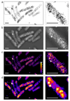
Super-Resolution Microscopy and Tracking of DNA-Binding Proteins in Bacterial Cells
- PMID: 27283312
- PMCID: PMC4970795
- DOI: 10.1007/978-1-4939-3631-1_16
Super-Resolution Microscopy and Tracking of DNA-Binding Proteins in Bacterial Cells
Abstract
The ability to detect individual fluorescent molecules inside living cells has enabled a range of powerful microscopy techniques that resolve biological processes on the molecular scale. These methods have also transformed the study of bacterial cell biology, which was previously obstructed by the limited spatial resolution of conventional microscopy. In the case of DNA-binding proteins, super-resolution microscopy can visualize the detailed spatial organization of DNA replication, transcription, and repair processes by reconstructing a map of single-molecule localizations. Furthermore, DNA-binding activities can be observed directly by tracking protein movement in real time. This allows identifying subpopulations of DNA-bound and diffusing proteins, and can be used to measure DNA-binding times in vivo. This chapter provides a detailed protocol for super-resolution microscopy and tracking of DNA-binding proteins in Escherichia coli cells. The protocol covers the construction of cell strains and describes data acquisition and analysis procedures, such as super-resolution image reconstruction, mapping single-molecule tracks, computing diffusion coefficients to identify molecular subpopulations with different mobility, and analysis of DNA-binding kinetics. While the focus is on the study of bacterial chromosome biology, these approaches are generally applicable to other molecular processes and cell types.
Keywords: DNA repair; DNA-binding proteins; Escherichia coli; Lambda red recombination; Single-molecule imaging; Single-particle tracking; Super-resolution fluorescence microscopy.
Figures


Similar articles
-
Super-Resolution Microscopy and Tracking of DNA-Binding Proteins in Bacterial Cells.Methods Mol Biol. 2022;2476:191-208. doi: 10.1007/978-1-0716-2221-6_15. Methods Mol Biol. 2022. PMID: 35635706
-
Bound2Learn: a machine learning approach for classification of DNA-bound proteins from single-molecule tracking experiments.Nucleic Acids Res. 2021 Aug 20;49(14):e79. doi: 10.1093/nar/gkab186. Nucleic Acids Res. 2021. PMID: 33744965 Free PMC article.
-
Single-molecule and super-resolution imaging of transcription in living bacteria.Methods. 2017 May 1;120:103-114. doi: 10.1016/j.ymeth.2017.04.001. Epub 2017 Apr 13. Methods. 2017. PMID: 28414097 Free PMC article. Review.
-
SMTracker: a tool for quantitative analysis, exploration and visualization of single-molecule tracking data reveals highly dynamic binding of B. subtilis global repressor AbrB throughout the genome.Sci Rep. 2018 Oct 24;8(1):15747. doi: 10.1038/s41598-018-33842-9. Sci Rep. 2018. PMID: 30356068 Free PMC article.
-
In vivo single-molecule imaging of bacterial DNA replication, transcription, and repair.FEBS Lett. 2014 Oct 1;588(19):3585-94. doi: 10.1016/j.febslet.2014.05.026. Epub 2014 May 23. FEBS Lett. 2014. PMID: 24859634 Review.
Cited by
-
ExTrack characterizes transition kinetics and diffusion in noisy single-particle tracks.J Cell Biol. 2023 May 1;222(5):e202208059. doi: 10.1083/jcb.202208059. Epub 2023 Feb 28. J Cell Biol. 2023. PMID: 36880553 Free PMC article.
-
Alteration of DNA supercoiling serves as a trigger of short-term cold shock repressed genes of E. coli.Nucleic Acids Res. 2022 Aug 26;50(15):8512-8528. doi: 10.1093/nar/gkac643. Nucleic Acids Res. 2022. PMID: 35920318 Free PMC article.
-
Phenotypic heterogeneity in the bacterial oxidative stress response is driven by cell-cell interactions.Cell Rep. 2023 Mar 28;42(3):112168. doi: 10.1016/j.celrep.2023.112168. Epub 2023 Feb 26. Cell Rep. 2023. PMID: 36848288 Free PMC article.
-
Transient non-specific DNA binding dominates the target search of bacterial DNA-binding proteins.Mol Cell. 2021 Apr 1;81(7):1499-1514.e6. doi: 10.1016/j.molcel.2021.01.039. Epub 2021 Feb 22. Mol Cell. 2021. PMID: 33621478 Free PMC article.
References
MeSH terms
Substances
Grants and funding
LinkOut - more resources
Full Text Sources
Other Literature Sources

