. 2016 Jun 10:6:27539.
doi: 10.1038/srep27539.

Curcumin inhibits HIV-1 by promoting Tat protein degradation

Affiliations

Curcumin inhibits HIV-1 by promoting Tat protein degradation

Amjad Ali et al. Sci Rep. .

Abstract

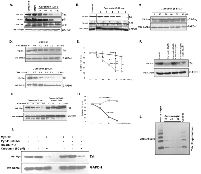
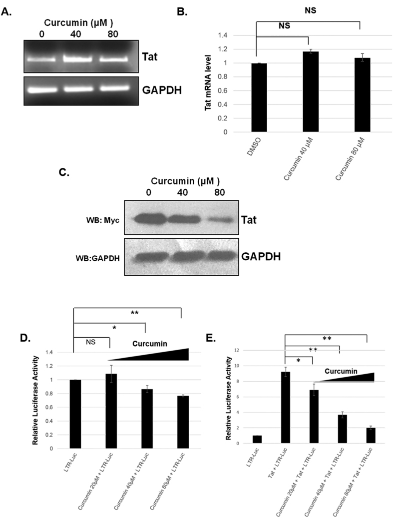
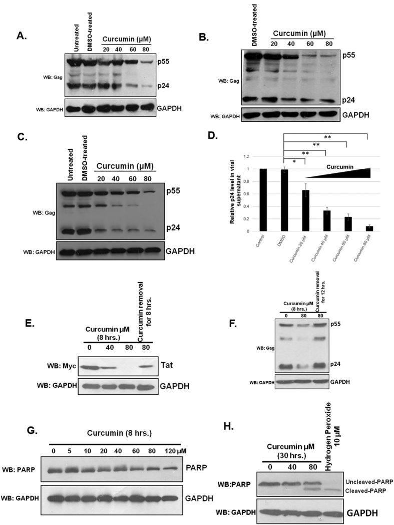
HIV-1 Tat is an intrinsically unfolded protein playing a pivotal role in viral replication by associating with TAR region of viral LTR. Unfolded proteins are degraded by 20S proteasome in an ubiquitin independent manner. Curcumin is known to activate 20S proteasome and promotes the degradation of intrinsically unfolded p53 tumor suppressor protein. Since HIV-1 Tat protein is largerly unfolded, we hypothesized that Tat may also be targeted through this pathway. Curcumin treated Tat transfected HEK-293T cells showed a dose and time dependent degradation of Tat protein. Contrary to this HIV-1 Gag which is a properly folded protein, remained unaffected with curcumin. Semi-quantitative RT-PCR analysis showed that curcumin treatment did not affect Tat gene transcription. Curcumin increased the rate of Tat protein degradation as shown by cycloheximide (CHX) chase assay. Degradation of the Tat protein is accomplished through proteasomal pathway as proteasomal inhibitor MG132 blocked Tat degradation. Curcumin also decreased Tat mediated LTR promoter transactivation and inhibited virus production from HIV-1 infected cells. Taken together our study reveals a novel observation that curcumin causes potent degradation of Tat which may be one of the major mechanisms behind its anti HIV activity.


Figures

Figure 1
Figure 1. Curcumin decreased HIV-1 Tat protein.
(A) HEK-293T cells were transfected with 1 μg of Myc-Tat expressing plasmid, and after 36 hrs treated with curcumin for 8 hrs, lysed and probed for Tat, p53 and GAPDH. pEGFP-N1 (50 ng) was also transfected as transfection control. The blot shown is a representative of three independent experiments.(B) Myc-Tat was transfected in HEK-293T cells and curcumin treatment was performed for increasing time period. The blot is a representative of three independent experiments. (C) Gag-Opt (1 μg) was transfected and curcumin treatment was performed followed by immmuno-blotting for Gag protein in HHEK-293T cells. (D) Myc-Tat transfected HEK-293T cells were treated with CHX alone or with curcumin for time periods as indicated and Tat protein level was measured by western blotting. (E) The mean value of Tat protein from three independent experiments was plotted with respect to treatment period. P value was calculated by a two-tailed t-test (*P < 0.05, **P < 0.01; NS, not significant). (F) The Myc-Tat transfected HEK-293T cells were treated with curcumin, along with proteasomal and lysosomal inhibitors MG132 and ammonium chloride for 8 hrs, subsequently Tat level was measured. (G) Myc-Tat transfected HEK-293T cells were treated with curcumin and CHX in the absence or presence of MG132 for different time periods followed by western blotting for Tat protein. (H) Densitometry of Tat bands was carried out by using image J and plotted with respect to treatment period. (I) HEK-293T cells were transfected with 1 μg of Myc-Tat (lanes 1–3) for 36 hrs followed by treatment with curcumin and Pyr-41 for 6 hrs subsequently the western blotting was done for Tat protein. HEK-293T cells were transfected with 1 μg of Myc-Tat and 2 μg of HA-Ub KO, 36 hrs of transfection curcumin treatment was done for 6 hrs and Tat protein was blotted. (J) HEK-293T cells were transfected with 1 μg of Myc-Tat and 2 μg of 6X-His Ubiquitin plasmid, after 36 hrs the cells were treated with increasing dose of curcumin or MG132. The ubiquitinated proteins were purified using Ni-NTA affinity chromatography and ubiquitinated Tat was blotted using anti-Myc antibody. The original uncropped blot images for (A–D,F,G,I,J) are shown in supplementary information as supplementary figure.
Figure 2
Figure 2. Curcumin does not modulate the level of Tat mRNA.
(A) Myc-Tat transfected HEK-293T cells were treated with curcumin for 8 hrs and total RNA was isolated using TRIZOL reagent followed by RT-PCR using Tat and GAPDH primers. The gel image is a representative of three independent experiments. (B) Using ImageJ the band intensity of Tat and GAPDH was quantified and plotted as Tat/GAPDH. P value was calculated by a two-tailed t-test (*P < 0.05, **P < 0.01; NS, not significant). (C) Myc-Tat was transfected to HEK-293T cells, 24 hrs post transfection the cell culture medium was applied on fresh HEK-293T cells followed by treatment with curcumin for 6 hrs. Western blotting was done to detect the Tat protein. (D) In a 24 well plate format TZM-bl cells were transfected with 0.2 μg of Myc Tat, after 36 hrs treated with increasing dose of curcumin for 12 hrs and luciferase activity was measured and the mean of three independent experiments was plotted. P value was calculated by a two-tailed t-test (*P < 0.05, **P < 0.01; NS, not significant). (E) Similarly un-transfected TZM-bl cells were also treated with curcumin and luciferase activity was measured and the mean of three independent experiments was plotted. P value was calculated by a two-tailed t-test (*P < 0.05, **P < 0.01; NS, not significant).
Figure 3
Figure 3. Curcumin treatment resulted in a reduced production of HIV-1 virions.
(A) HEK-293T cells were transfected with 1 μg pNL4-3, after 36 hrs the medium was changed and curcumin was added from 20–80 μM for 12 hrs. The cells were lysed and probed for p24 Gag protein. Medium containing viral supernatant was used to infect TZM-bl cells. (B) TZM-bl cells were infected with viral supernatant obtained from previous experiment and the cells were further incubated for 24 hrs followed by lysis and measurement of p24 level. (C) J1.1 cells were stimulated with 20 ng/ml of TNF-α for 12 hrs subsequently the medium was replaced with fresh complete RPMI containing curcumin and TNF-α as indicated. The cells were lysed and blotted for p24 level whereas the cell culture medium containing virions were used to detect viral load by direct ELISA. (D) The virus containing medium from previous experiment was coated on 96 well ELISA plate and probed with p24 antibody, the average value of p24 from three independent ELISA experiments was plotted with respect to curcumin concentration. P value was calculated by a two-tailed t-test (*P < 0.05, **P < 0.01; NS, not significant). (E) Myc-Tat transfected HEK-293T cells were treated with curcumin for 8 hrs subsequently it was removed and fresh complete DMEM was added and incubated for 8 hrs followed by immunoblotting for Tat. (F) Curcumin treatment was carried out to pNL4-3 transfected HEK-293T cells followed by replacement with fresh DMEM medium and further incubation for 12 hrs. The p24 protein level was measured by western blotting. (G) HEK-293T cells were treated with indicated doses of curcumin for 8 hrs followed by western blotting for PARP. (H) Curcumin treatment was carried out for 30 hrs followed by western blotting to detect un-cleaved and cleaved PARP. As a control the cells were treated with hydrogen peroxide and blotted for PARP.

References

    1. Ciechanover A. Intracellular protein degradation: from a vague idea thru the lysosome and the ubiquitin-proteasome system and onto human diseases and drug targeting. Cell Death Differ. 12, 1178–1190 (2005). - PubMed
    1. Hochstrasser M. Ubiquitin-dependent protein degradation. Annu. Rev. Genet. 30, 405–439 (1996). - PubMed
    1. Voges D., Zwickl P. & Baumeister W. The 26S proteasome: a molecular machine designed for controlled proteolysis. Annu. Rev. Biochem. 68, 1015–1068 (1999). - PubMed
    1. Asher G., Reuven N. & Shaul Y. 20S proteasomes and protein degradation “by default”. Bioessays 28, 844–849 (2006). - PubMed
    1. Jariel-Encontre I., Bossis G. & Piechaczyk M. Ubiquitin-independent degradation of proteins by the proteasome. Biochim. Biophys. Acta 1786, 153–177 (2008). - PubMed

Publication types

MeSH terms

Substances