

Crystal structure of NOD2 and its implications in human disease
- PMID: 27283905
- PMCID: PMC4906405
- DOI: 10.1038/ncomms11813
Crystal structure of NOD2 and its implications in human disease
Abstract
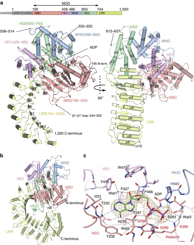
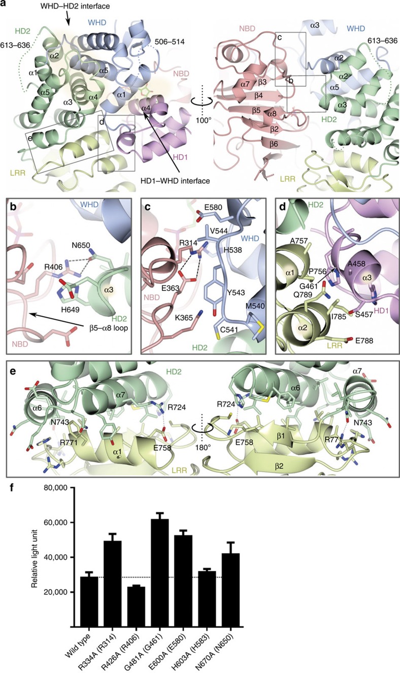
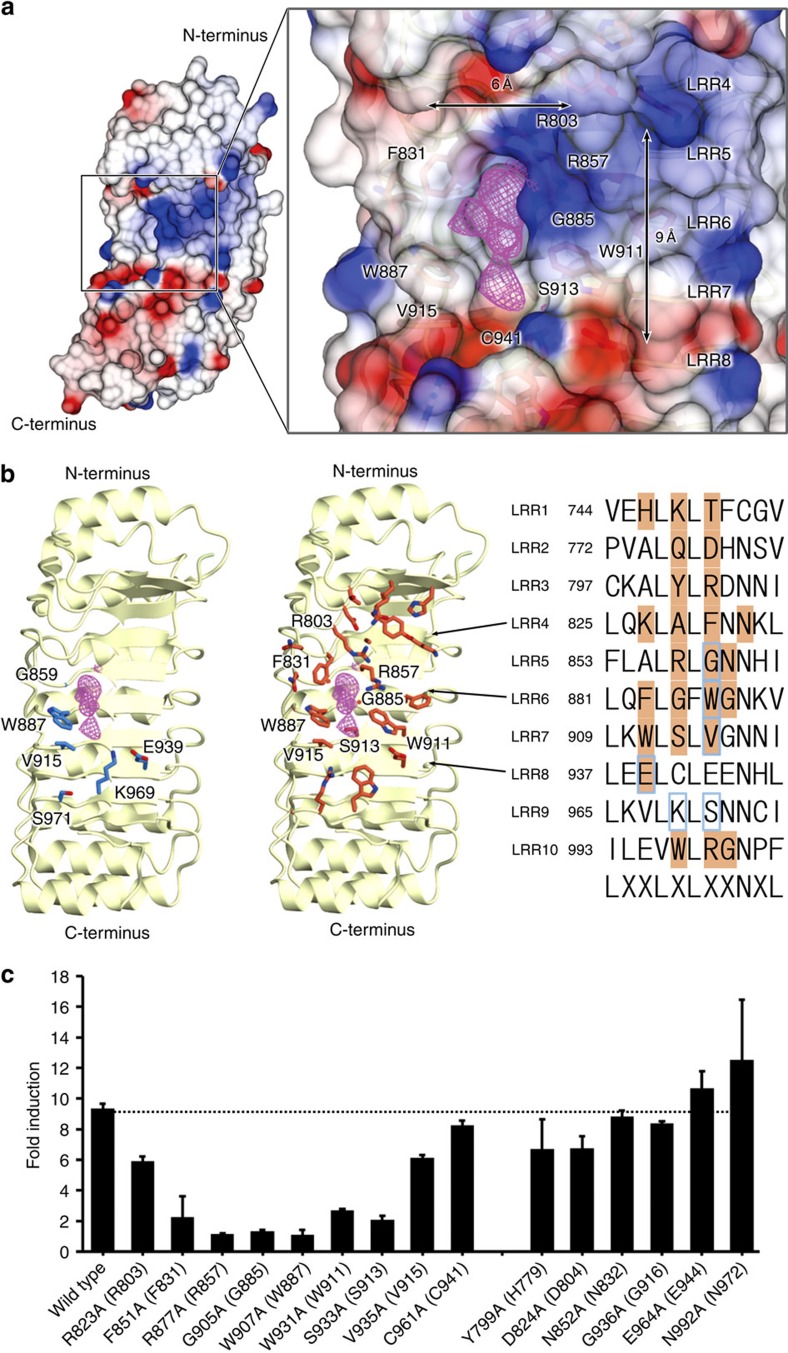
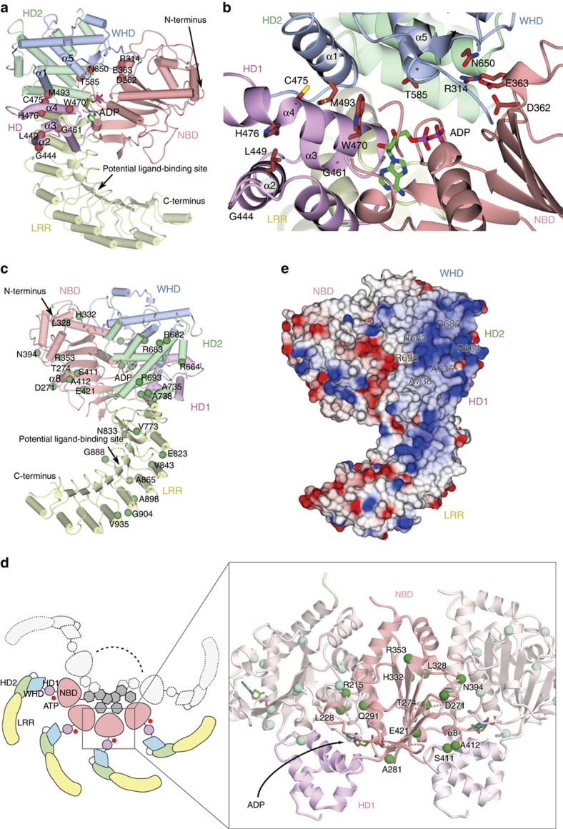
Nucleotide-binding oligomerization domain-containing protein 2 (NOD2), a member of the NOD-like receptors family, are crucial for innate immune responses. Mutations of NOD2 have been associated with chronic inflammatory disorders such as Crohn's disease (CD), Blau syndrome (BS) and early-onset sarcoidosis (EOS), but little is known about its signalling mechanism and the role it plays in these diseases. Here, we report the crystal structure of rabbit NOD2 in an ADP-bound state. The structure reveals an inactive closed conformation in which the subdomains in the NOD domain are closely packed by ADP-mediated and inter-domain interactions. Mapping of the BS- or EOS-associated gain-of-function mutations reveals that most of these mutations are located in the NOD subdomain interfaces, and are likely to disrupt the inner domain interactions, facilitating a conformational change to the active form. Conversely, mutations associated with CD are distributed throughout the protein, some of which may affect oligomer formation and ligand binding.
Figures
Similar articles
-
Computational model to analyze and characterize the functional mutations of NOD2 protein causing inflammatory disorder - Blau syndrome.Adv Protein Chem Struct Biol. 2020;120:379-408. doi: 10.1016/bs.apcsb.2019.11.005. Epub 2020 Feb 4. Adv Protein Chem Struct Biol. 2020. PMID: 32085886 Review.
-
Structural models of zebrafish (Danio rerio) NOD1 and NOD2 NACHT domains suggest differential ATP binding orientations: insights from computational modeling, docking and molecular dynamics simulations.PLoS One. 2015 Mar 26;10(3):e0121415. doi: 10.1371/journal.pone.0121415. eCollection 2015. PLoS One. 2015. PMID: 25811192 Free PMC article.
-
Nod2-Nodosome in a Cell-Free System: Implications in Pathogenesis and Drug Discovery for Blau Syndrome and Early-Onset Sarcoidosis.ScientificWorldJournal. 2016;2016:2597376. doi: 10.1155/2016/2597376. Epub 2016 Jun 15. ScientificWorldJournal. 2016. PMID: 27403452 Free PMC article.
-
Nucleotide-binding oligomerization domain containing 2: structure, function, and diseases.Semin Arthritis Rheum. 2013 Aug;43(1):125-30. doi: 10.1016/j.semarthrit.2012.12.005. Epub 2013 Jan 24. Semin Arthritis Rheum. 2013. PMID: 23352252 Review.
-
Computational studies on receptor-ligand interactions between novel buffalo (Bubalus bubalis) nucleotide-binding oligomerization domain-containing protein 2 (NOD2) variants and muramyl dipeptide (MDP).J Mol Graph Model. 2016 Apr;65:15-26. doi: 10.1016/j.jmgm.2016.02.004. Epub 2016 Feb 10. J Mol Graph Model. 2016. PMID: 26897084
Cited by
-
Ripks and Neuroinflammation.Mol Neurobiol. 2024 Sep;61(9):6771-6787. doi: 10.1007/s12035-024-03981-4. Epub 2024 Feb 13. Mol Neurobiol. 2024. PMID: 38349514 Review.
-
Designed leucine-rich repeat proteins bind two muramyl dipeptide ligands.Protein Sci. 2021 Apr;30(4):804-817. doi: 10.1002/pro.4031. Epub 2021 Feb 15. Protein Sci. 2021. PMID: 33512005 Free PMC article.
-
RIP2 filament formation is required for NOD2 dependent NF-κB signalling.Nat Commun. 2018 Oct 2;9(1):4043. doi: 10.1038/s41467-018-06451-3. Nat Commun. 2018. PMID: 30279485 Free PMC article.
-
A Novel Rare Missense Variation of the NOD2 Gene: Evidencesof Implication in Crohn's Disease.Int J Mol Sci. 2019 Feb 15;20(4):835. doi: 10.3390/ijms20040835. Int J Mol Sci. 2019. PMID: 30769939 Free PMC article.
-
Double autoinhibition mechanism of signal transduction ATPases with numerous domains (STAND) with a tetratricopeptide repeat sensor.Nucleic Acids Res. 2019 Apr 23;47(7):3795-3810. doi: 10.1093/nar/gkz112. Nucleic Acids Res. 2019. PMID: 30788511 Free PMC article.
References
-
- Lamkanfi M. & Dixit V. M. Mechanisms and functions of inflammasomes. Cell 157, 1013–1022 (2014). - PubMed
-
- Schroder K. & Tschopp J. The inflammasomes. Cell 140, 821–832 (2010). - PubMed
-
- Takeuchi O. & Akira S. Pattern recognition receptors and inflammation. Cell 140, 805–820 (2010). - PubMed
-
- von Moltke J., Ayres J. S., Kofoed E. M., Chavarria-Smith J. & Vance R. E. Recognition of bacteria by inflammasomes. Annu. Rev. Immunol. 31, 73–106 (2013). - PubMed
-
- Danot O., Marquenet E., Vidal-Ingigliardi D. & Richet E. Wheel of life, wheel of death: a mechanistic insight into signaling by STAND proteins. Structure 17, 172–182 (2009). - PubMed
Publication types
MeSH terms
Substances
LinkOut - more resources
Full Text Sources
Other Literature Sources
Molecular Biology Databases
