. 2016 Aug 5;291(32):16879-91.
doi: 10.1074/jbc.M116.728600. Epub 2016 Jun 9.

Dual Function of Phosphoubiquitin in E3 Activation of Parkin

Affiliations

Dual Function of Phosphoubiquitin in E3 Activation of Parkin

Erik Walinda et al. J Biol Chem. .

Abstract

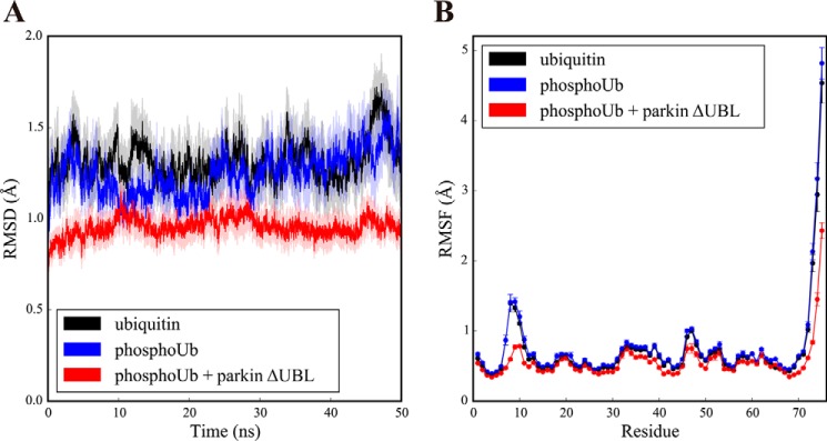
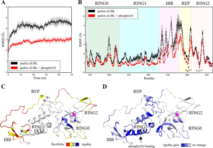
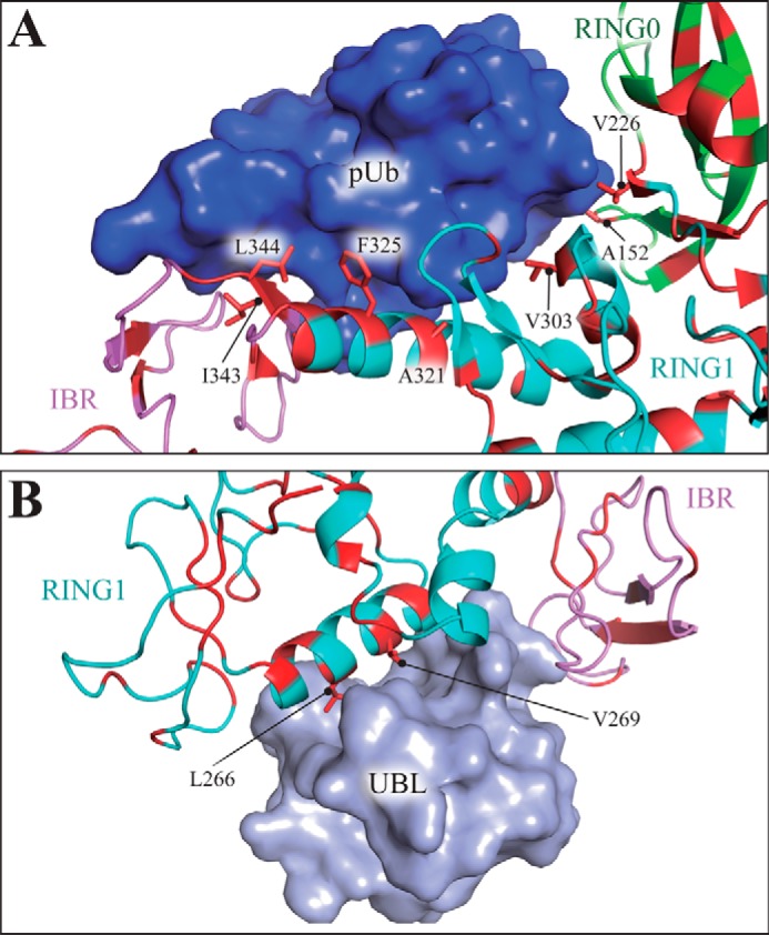
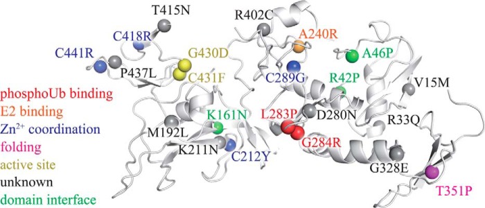
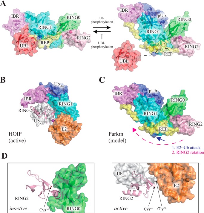
Mutations in the gene encoding parkin, an auto-inhibited E3 ubiquitin ligase that functions in the clearance of damaged mitochondria, are the most common cause of autosomal recessive juvenile Parkinsonism. The mechanism regulating parkin activation remains poorly understood. Here we show, by using isothermal titration calorimetry, solution NMR, and fluorescence spectroscopy, that parkin can bind ubiquitin and phosphomimetic ubiquitin by recognizing the canonical hydrophobic patch and C terminus of ubiquitin. The affinity of parkin for both phosphomimetic and unmodified ubiquitin is markedly enhanced upon removal of the ubiquitin-like (UBL) domain of parkin. This suggests that the agonistic binding of ubiquitin to parkin in trans is counterbalanced by the antagonistic activity of the parkin UBL domain in cis Intriguingly, UBL binding is enthalpy-driven, whereas ubiquitin binding is driven by an increase in the total entropy of the system. These thermodynamic differences are explained by different chemistry in the ubiquitin- and UBL-binding pockets of parkin and, as shown by molecular dynamics simulations, are not a consequence of changes in protein conformational entropy. Indeed, comparison of conformational fluctuations reveals that the RING1-IBR element becomes considerably more rigid upon complex formation. A model of parkin activation is proposed in which E2∼Ub binding triggers large scale diffusional motion of the RING2 domain toward the ubiquitin-stabilized RING1-IBR assembly to complete formation of the active parkin-E2∼Ub transfer complex. Thus, ubiquitin plays a dual role in parkin activation by competing with the inhibitory UBL domain and stabilizing the active form of parkin.

Keywords: Parkinson disease; isothermal titration calorimetry (ITC); molecular dynamics; parkin; protein dynamics; protein-protein interaction; ubiquitin; ubiquitin ligase.


Figures

FIGURE 1.
FIGURE 1.
Parkin is an auto-inhibited E3 enzyme that requires structural rearrangement for activation. A, domain architecture of rat parkin, comprising a UBL domain, an ∼70-residue linker, and four domains of the RING family. Parkin is an RBR-type ubiquitin ligase with its catalytic center embedded in the most C-terminal RING2 domain (Cys431 in rat parkin). The RING1 domain serves as an adapter platform for the incoming E2 enzyme. The yellow region represents a short REP, which functions in the auto-inhibition of parkin (7). B, quaternary structure of full-length parkin, drawn from PDB code 4K95 (7). Key sites are indicated. Important auto-inhibitory interactions in parkin include blockade of the catalytic cysteine by the RING0 domain and occlusion of the E2-binding site (blue) by the REP element. C and D, structural comparison of free parkin (C) from R. norvegicus (PDB code 4K95 (7)) and phosphoubiquitin-bound parkin (D) from P. humanus (PDB code 5CAW (19)). In both crystal structures, the catalytic cysteine is occluded by the RING0 domain. Protein representations were generated by PyMOL (Schrödinger, LLC.).
FIGURE 2.
FIGURE 2.
Physical interaction of parkin with ubiquitin. A, transverse relaxation-optimized spectroscopy-HSQC spectrum of 100 μm 15N-labeled ubiquitin before (black) and after (red) addition of 1 mol eq of unlabeled full-length parkin, showing selective line broadening after the addition of parkin. B, residue-specific NMR signal loss in ubiquitin upon the addition of parkin. Green bars, residues not observed in the reference spectrum of free ubiquitin; red bars, residues for which near-complete signal loss occurred upon the addition of parkin. Red line, average intensity ratio. C, structural model of ubiquitin binding to parkin based on PDB code 5CAW (19). Residues (one-letter code) with significant line broadening in the NMR experiment are colored red.
FIGURE 3.
FIGURE 3.
Ubiquitin binding to parkin is entropy-driven. A and B, isothermal titration calorimetry thermogram for ubiquitin (A) and phosphomimetic ubiquitin (B) binding to full-length parkin, showing an endothermic reaction. Upper panels show raw data; lower panels show the integrated heat values. C, dissociation constants determined from the ITC experiments. Unmodified ubiquitin and phosphomimetic ubiquitin bind to full-length parkin with similar avidity (p = 0.16; Student's t test). D, entropy drives the binding of both ubiquitin and phosphomimetic ubiquitin to parkin. The binding process is disfavored enthalpically, but this is counteracted by a large favorable entropic term. Error bars indicate the S.E. in C and D of three independent experiments (n = 3).
FIGURE 4.
FIGURE 4.
Binding of ubiquitin, phosphomimetic ubiquitin, and parkin UBL to the parkin core (parkin ΔUBL). A–C, isothermal titration calorimetry thermograms for ubiquitin (A), phosphomimetic ubiquitin (B), and parkin UBL (C) binding to parkin ΔUBL. Upper panels, raw data; lower panels, integrated heat values. D, dissociation constants determined from the ITC experiments. Phosphomimetic ubiquitin binds to the parkin core more avidly than wild-type ubiquitin (p < 0.001; Student's t test). E, thermodynamic parameters for the binding of ubiquitin, phosphomimetic ubiquitin, and the parkin UBL to parkin ΔUBL. Error bars indicate the S.E. in D and E of three independent experiments (n = 3).
FIGURE 5.
FIGURE 5.
Structural changes in the parkin core upon ubiquitin binding. A, distribution of tryptophan residues in rat parkin. Pink sphere, catalytic cysteine. B, changes in the tryptophan emission spectrum of full-length parkin upon binding to ubiquitin. C, red shift of the baricentric mean of the fluorescence emission spectrum as a function of the molar ratio of ubiquitin to parkin. D, changes in the tryptophan emission spectrum of parkin ΔUBL upon binding to ubiquitin. a.u., arbitrary unit. E, red shift of the baricentric mean of the fluorescence emission spectrum as a function of the molar ratio of ubiquitin to parkin ΔUBL.
FIGURE 6.
FIGURE 6.
Changes in conformational fluctuations in ubiquitin upon binding to the parkin core. A, time traces of the r.m.s.d. calculated for Cα atoms after rotational and translational fitting to the Cα coordinates of the first time frame during molecular dynamics simulations of ubiquitin (black), phosphoubiquitin (blue), and phosphoubiquitin in the parkin-phosphoubiquitin complex (red). All simulations were performed 10 times with different randomized initial velocities. Average r.m.s.d. values are shown. The shaded area represents the S.E. of the respective r.m.s.d. B, root mean square fluctuation (RMSF) of Cα atoms of ubiquitin (black), phosphoubiquitin (blue), and phosphoubiquitin in the parkin-phosphoubiquitin complex (red) averaged over the course of the simulations. The average RMSF value of 10 simulations, each of 50 ns, is shown. Error bars indicate the S.E.
FIGURE 7.
FIGURE 7.
Conformational fluctuations in the parkin core before and after binding to phosphoubiquitin. A, time traces of the r.m.s.d. calculated for Cα atoms after rotational and translational fitting to the Cα coordinates of the first time frame during molecular dynamics simulations of parkin ΔUBL (black) and the parkin ΔUBL-phosphoubiquitin complex (red). All simulations were performed 10 times with different randomized initial velocities. Average r.m.s.d. values are shown. The shaded area represents the S.E. of the respective r.m.s.d. B, RMSF of Cα atoms of parkin ΔUBL (black) and the parkin ΔUBL-phosphoubiquitin complex (red) averaged over the time interval (5 ns; 50 ns). The average RMSF value of 10 simulations, each of 50 ns, is shown. Error bars indicate the S.E. Residue numbers refer to parkin from R. norvegicus. and the alignment was performed by Clustal Omega (53). The respective domains are indicated. C, conformational fluctuations in the free parkin core (B) as visualized on the structure of the first time frame. Residues are color-coded as follows: red, RMSF > RMSFaverage + 1σ; yellow, RMSF > RMSFaverage; blue, RMSF < RMSFaverage − 1σ; gray, all other residues. Magenta sphere, catalytic cysteine. D, changes in conformational fluctuations in the parkin core upon binding to phosphoubiquitin. The ratio of the RMSF values of B is visualized on the structure of free parkin. The phosphoubiquitin-binding site is indicated. Color code: blue, ρi > ρaverage + 1σ; light blue, ρi > ρaverage; gray, all other residues; ρi, RMSF (B) ratio for residue i of the free to the phosphoubiquitin-bound form of the parkin core. Magenta sphere, catalytic cysteine.
FIGURE 8.
FIGURE 8.
Distinct properties of the UBL- and phosphoubiquitin-binding sites on parkin. A, binding site of phosphoubiquitin on parkin contains several hydrophobic residues (red), many of which engage in interactions with phosphoubiquitin, thereby requiring solvent exposure before complex formation. B, conversely, only two hydrophobic side chains of the parkin core engage in interactions with the UBL domain of parkin. The structures are drawn from PDB code 5CAV (A) (19) and 4K95 (B) (7). Color code: RING0, green; RING1, cyan; IBR, violet; phosphoubiquitin, dark blue; UBL, light blue; residues with exposed hydrophobic side chains (Phe, Val, Leu, Ile, Trp, Met, and Ala) are shown in red.
FIGURE 9.
FIGURE 9.
AR-JP mutations may unbalance the conformational dynamics of parkin by a variety of mechanisms. Pathogenic mutations linked to autosomal recessive juvenile parkinsonism (3, 30, 54–62) are mapped onto the crystal structure of parkin (spheres). Several mutants appear to affect the structure of parkin by interfering with Zn2+ binding (blue) or the correct formation of secondary structure elements (magenta). The mutants G430D and C431F directly affect the active site (yellow). Other PD mutants may alter domain-domain interactions (green). Ala240 (orange) is critical for binding to the E2, and Leu283 and Gly284 (red) participate in phosphoubiquitin binding (19). Many of the remaining PD mutants are less well understood (gray) but may modulate the overall conformational dynamics of the enzyme. The structure of parkin was drawn from PDB code 4K95 (7).
FIGURE 10.
FIGURE 10.
Model of parkin activation by phosphoubiquitin. A, left, in the initial state, parkin adopts an auto-inhibited conformation with the UBL domain bound to the parkin core (7). PINK1 phosphorylates Ser65 of ubiquitin, which competes with the UBL for binding to the parkin core. Release (19, 29) of the UBL domain from the parkin core to the solvent enables PINK1 to phosphorylate Ser65 of the UBL, which prevents the UBL from rebinding back on the parkin core. Accordingly, the action of PINK1 drives the UBL/phosphoubiquitin competition equilibrium toward a phosphoubiquitin-bound/UBL-exposed state of parkin (right, schematic). B, structure of an active RBR/E2∼Ub transfer complex of the RBR-type E3 ubiquitin ligase HOIP (31). The linear ubiquitin chain-determining domain is omitted for clarity. An allosteric ubiquitin molecule (Uballo) orients the IBR and RING1 domains with respect to one other. C, as compared with the inactive conformation of parkin (A), the RING2 domain in active HOIP (B) is rotated and translated, bringing it in close proximity to the RING1-IBR element. Such a large scale structural change is permitted by the long linker between the IBR and RING2 domains, but it requires release of the REP element from RING1. This displacement of the REP element is probably driven by competitive binding of E2∼Ub to the E2-binding site on RING1 (blue arrow). D, comparison of the inactive and active states of the RING2 domain of parkin. Left, in the inactive conformation, the catalytic cysteine is occluded by the RING0 domain. Right, structural model of the RING2 domain of parkin in the active state as generated by SWISS-MODEL (63) on the basis of the active state of HOIP (B). The sequence identity of the RING2 domain between HOIP and parkin, as judged by SWISS-MODEL, is 43%. Apart from formation of a new α-helix, the overall structure of the RING2 domain is not markedly changed, indicating that simple rotation and translation of the RING2 domain with respect to the RING1-IBR element is sufficient to bring the catalytic cysteine within close proximity of the activated Gly76 of the incoming ubiquitin (Ubact), leading to formation of an active parkin-E2∼Ub transfer complex. Domains other than RING0 and RING2 are omitted for clarity. Structures are drawn from PDB codes 4K95, 5CAW, and 5EDV, respectively.

References

    1. Kitada T., Asakawa S., Hattori N., Matsumine H., Yamamura Y., Minoshima S., Yokochi M., Mizuno Y., and Shimizu N. (1998) Mutations in the parkin gene cause autosomal recessive juvenile parkinsonism. Nature 392, 605–608 - PubMed
    1. Shulman J. M., De Jager P. L., and Feany M. B. (2011) Parkinson's disease: genetics and pathogenesis. Annu. Rev. Pathol. 6, 193–222 - PubMed
    1. Lücking C. B., Dürr A., Bonifati V., Vaughan J., De Michele G., Gasser T., Harhangi B. S., Meco G., Denèfle P., Wood N. W., Agid Y., Brice A., French Parkinson's Disease Genetics Study Group, and European Consortium on Genetic Susceptibility in Parkinson's Disease (2000) Association between early-onset Parkinson's disease and mutations in the parkin gene. N. Engl. J. Med. 342, 1560–1567 - PubMed
    1. Wenzel D. M., Lissounov A., Brzovic P. S., and Klevit R. E. (2011) UBCH7 reactivity profile reveals parkin and HHARI to be RING/HECT hybrids. Nature 474, 105–108 - PMC - PubMed
    1. Matsuda N., Sato S., Shiba K., Okatsu K., Saisho K., Gautier C. A., Sou Y. S., Saiki S., Kawajiri S., Sato F., Kimura M., Komatsu M., Hattori N., and Tanaka K. (2010) PINK1 stabilized by mitochondrial depolarization recruits Parkin to damaged mitochondria and activates latent Parkin for mitophagy. J. Cell Biol. 189, 211–221 - PMC - PubMed

Publication types

MeSH terms

LinkOut - more resources