

Lysosomal and vacuolar sorting: not so different after all!
- PMID: 27284057
- PMCID: PMC5264500
- DOI: 10.1042/BST20160050
Lysosomal and vacuolar sorting: not so different after all!
Abstract
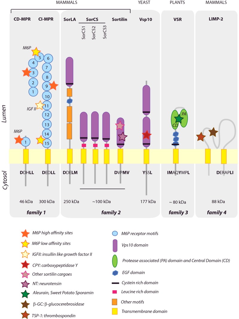
Soluble hydrolases represent the main proteins of lysosomes and vacuoles and are essential to sustain the lytic properties of these organelles typical for the eukaryotic organisms. The sorting of these proteins from ER residents and secreted proteins is controlled by highly specific receptors to avoid mislocalization and subsequent cellular damage. After binding their soluble cargo in the early stage of the secretory pathway, receptors rely on their own sorting signals to reach their target organelles for ligand delivery, and to recycle back for a new round of cargo recognition. Although signals in cargo and receptor molecules have been studied in human, yeast and plant model systems, common denominators and specific examples of diversification have not been systematically explored. This review aims to fill this niche by comparing the structure and the function of lysosomal/vacuolar sorting receptors (VSRs) from these three organisms.
Keywords: endosomal sorting; lysosomes; receptors; trafficking; vacuole.
© 2016 The Author(s). Published by Portland Press Limited on behalf of the Biochemical Society.
Figures
Similar articles
-
Sorting of plant vacuolar proteins is initiated in the ER.Plant J. 2010 May 1;62(4):601-14. doi: 10.1111/j.1365-313X.2010.04171.x. Epub 2010 Feb 10. Plant J. 2010. PMID: 20149141
-
A lysosomal biogenesis map reveals the cargo spectrum of yeast vacuolar protein targeting pathways.J Cell Biol. 2022 Apr 4;221(4):e202107148. doi: 10.1083/jcb.202107148. Epub 2022 Feb 17. J Cell Biol. 2022. PMID: 35175277 Free PMC article.
-
Trafficking of vacuolar proteins: the crucial role of Arabidopsis vacuolar protein sorting 29 in recycling vacuolar sorting receptor.Plant Cell. 2012 Dec;24(12):5058-73. doi: 10.1105/tpc.112.103481. Epub 2012 Dec 21. Plant Cell. 2012. PMID: 23263768 Free PMC article.
-
Plant RMR proteins: unique vacuolar sorting receptors that couple ligand sorting with membrane internalization.FEBS J. 2011 Jan;278(1):59-68. doi: 10.1111/j.1742-4658.2010.07923.x. Epub 2010 Nov 16. FEBS J. 2011. PMID: 21078125 Review.
-
Life and Death of Fungal Transporters under the Challenge of Polarity.Int J Mol Sci. 2020 Jul 29;21(15):5376. doi: 10.3390/ijms21155376. Int J Mol Sci. 2020. PMID: 32751072 Free PMC article. Review.
Cited by
-
Proteomic Analysis of Trichomonas vaginalis Phagolysosome, Lysosomal Targeting, and Unconventional Secretion of Cysteine Peptidases.Mol Cell Proteomics. 2022 Jan;21(1):100174. doi: 10.1016/j.mcpro.2021.100174. Epub 2021 Nov 8. Mol Cell Proteomics. 2022. PMID: 34763061 Free PMC article.
-
ReV as a Novel S. cerevisiae-Derived Drug Carrier to Enhance Anticancer Therapy through Daunorubicin Delivery.Appl Biochem Biotechnol. 2025 Apr;197(4):2301-2311. doi: 10.1007/s12010-024-05177-x. Epub 2024 Dec 30. Appl Biochem Biotechnol. 2025. PMID: 39738861
-
The Toxoplasma plant-like vacuolar compartment (PLVAC).J Eukaryot Microbiol. 2022 Nov;69(6):e12951. doi: 10.1111/jeu.12951. Epub 2022 Oct 27. J Eukaryot Microbiol. 2022. PMID: 36218001 Free PMC article. Review.
-
Protein Structure Insights into the Bilayer Interactions of the Saposin-Like Domain of Solanum tuberosum Aspartic Protease.Sci Rep. 2017 Dec 5;7(1):16911. doi: 10.1038/s41598-017-16734-2. Sci Rep. 2017. PMID: 29208977 Free PMC article.
-
A kinase cascade on the yeast lysosomal vacuole regulates its membrane dynamics: conserved kinase Env7 is phosphorylated by casein kinase Yck3.J Biol Chem. 2020 Aug 21;295(34):12262-12278. doi: 10.1074/jbc.RA119.012346. Epub 2020 Jul 9. J Biol Chem. 2020. PMID: 32647006 Free PMC article.
References
-
- van Leeuwenhoek A., Hoole S. The Select Works of Antony Van Leeuwenhoek, Containing His Microscopical Discoveries in Many of the Works of Nature. Translator; 1800:1800.
Publication types
MeSH terms
Substances
Grants and funding
LinkOut - more resources
Full Text Sources
Other Literature Sources
Molecular Biology Databases