. 2016 Apr 27;8(4):e588.
doi: 10.7759/cureus.588.

Management of Intracranial Meningiomas Using Keyhole Techniques

Affiliations

Management of Intracranial Meningiomas Using Keyhole Techniques

Joshua D Burks et al. Cureus. .

Abstract

Background: Keyhole craniotomies are increasingly being used for lesions of the skull base. Here we review our recent experience with these approaches for resection of intracranial meningiomas.

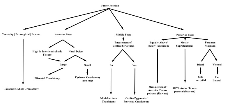
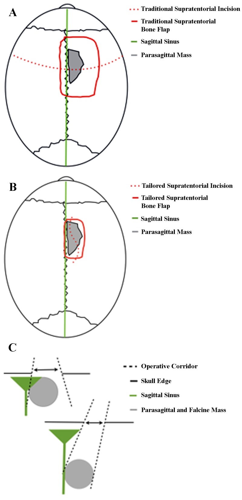
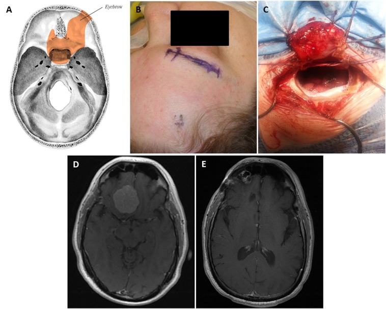
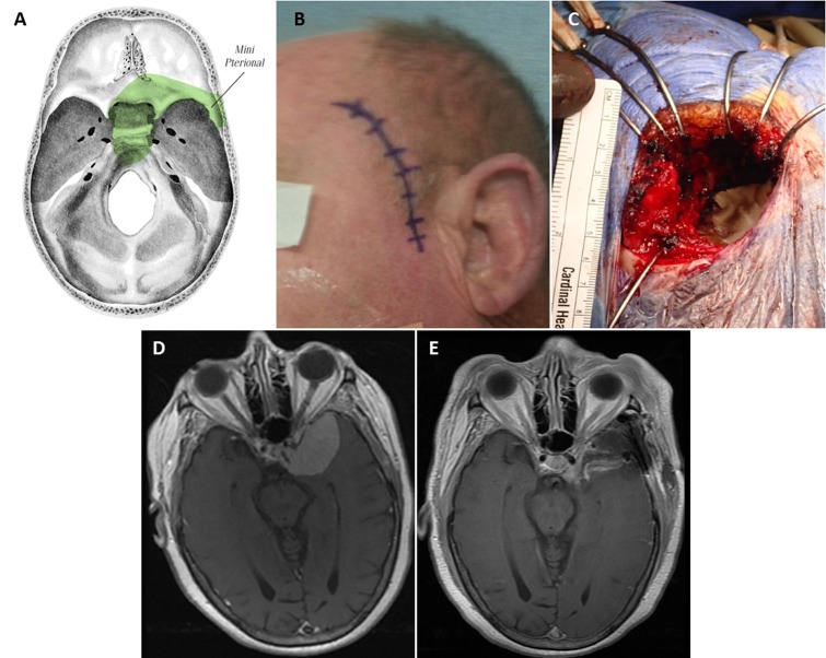
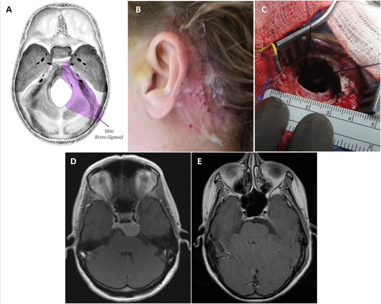
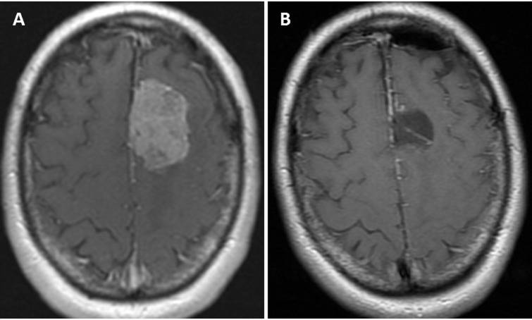
Methods: Clinical and operative data were gathered on all patients treated with keyhole approaches by the senior author from January 2012 to June 2013. Thirty-one meningiomas were resected in 27 patients, including 9 supratentorial, 5 anterior fossa, 7 middle fossa, 6 posterior fossa, and 4 complex skull base tumors. Twenty-nine tumors were WHO Grade I, and 2 were Grade II.

Results: The mean operative time was 8 hours, 22 minutes (range, 2:55-16:14) for skull-base tumors, and 4 hours, 27 minutes (range, 1:45-7:13) for supratentorial tumors. Simpson Resection grades were as follows: Grade I = 8, II = 8, III = 1, IV = 15, V = 0. The median postoperative hospital stay was 4 days (range, 1-20 days). In the 9 patients presenting with some degree of visual loss, 7 saw improvement or complete resolution. In the 6 patients presenting with cranial nerve palsies, 4 experienced improvement or resolution of the deficit postoperatively. Four patients experienced new neurologic deficits, all of which were improved or resolved at the time of the last follow-up. Technical aspects and surgical nuances of these approaches for management of intracranial meningiomas are discussed.

Conclusions: With careful preoperative evaluation, keyhole approaches can be utilized singly or in combination to manage meningiomas in a wide variety of locations with satisfactory results.

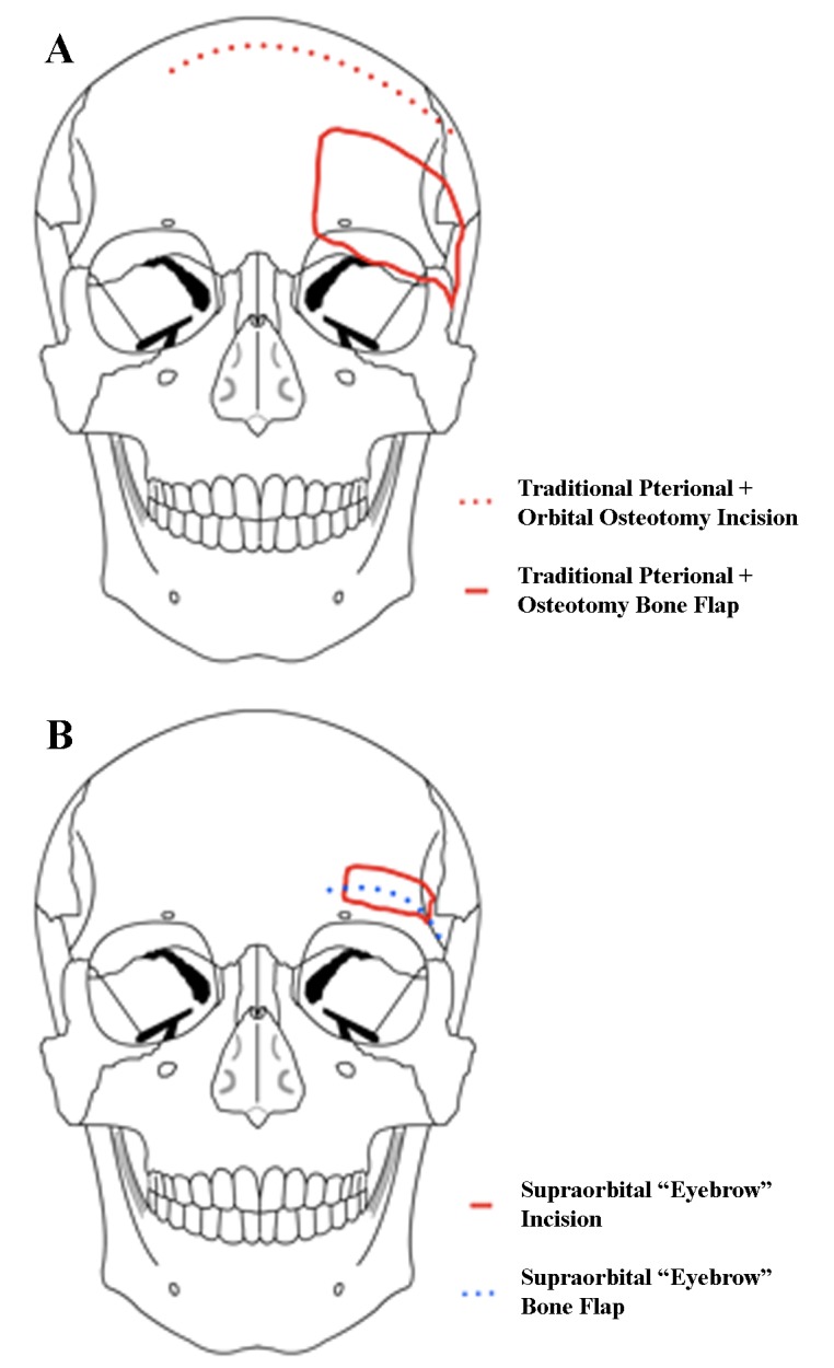
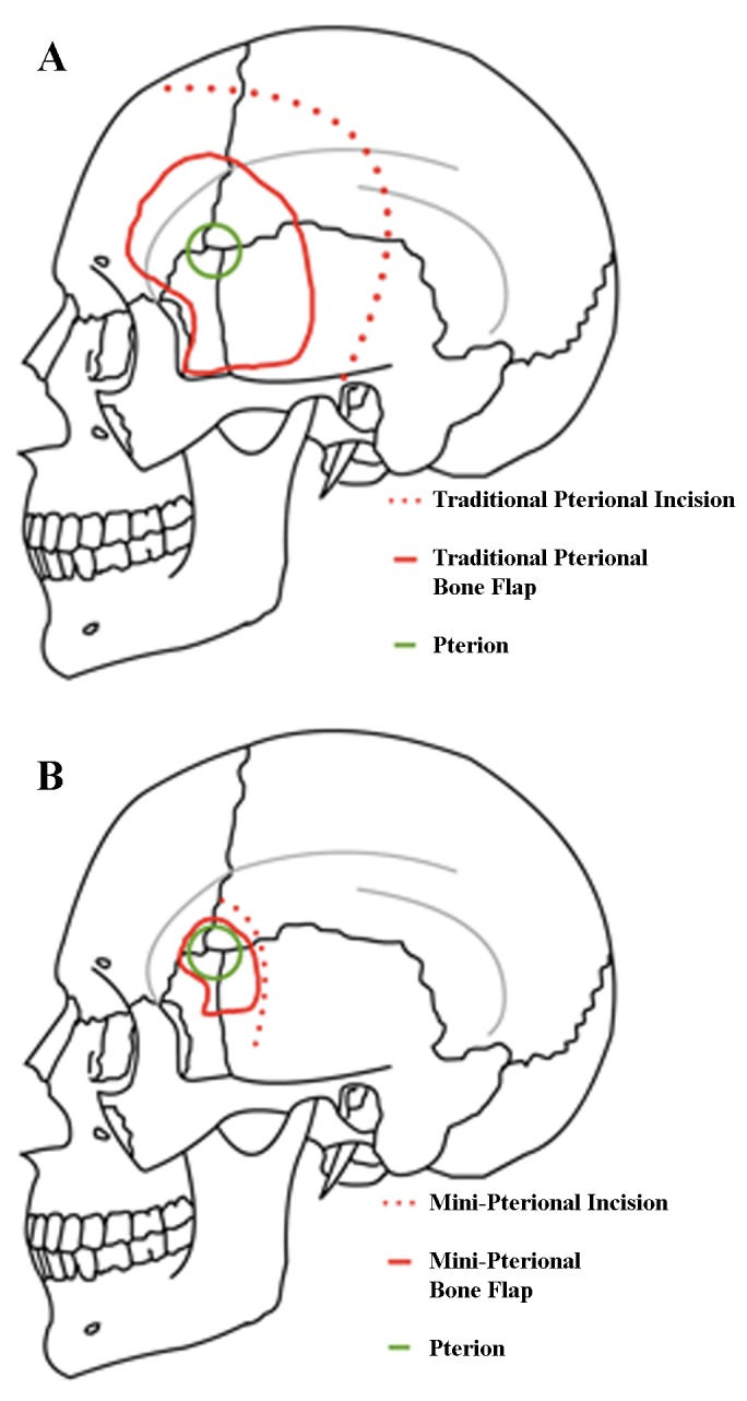
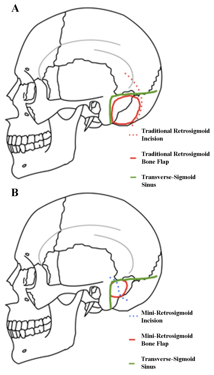
Keywords: craniotomy; eyebrow; keyhole; meningioma; pterional; retrosigmoid; supraorbital; tumor resection.


Conflict of interest statement

The authors have declared that no competing interests exist.

Figures

Figure 1
Figure 1. Our treatment paradigm for new meningiomas
Figure 2
Figure 2. Standard pterional and orbital osteotomy approach (A) vs. supra-orbital approach (B)
In a supraorbital approach, an incision is made within the eyebrow and lateral to the supraorbital nerve, and the frontalis muscle is divided parallel to the orbital rim and reflected downward. The keyhole is made just posterior to the temporal line, and the orbital rim is drilled flush with the orbital roof. Modified from: Rosario Van Tulpe, https://commons.wikimedia.org/wiki/File:SkullSchaedel3.png
Figure 3
Figure 3. Standard pterional approach (A) vs. mini-pterionaly approach (B)
The incision of a mini-pterional approach is made approximately 1 cm superior to the zygomatic arch and 1 cm anterior to the external auditory meatus, curving around the normal hairline and ending at the midline. The scalp flap and superficial fat pad are elevated together and reflected anteriorly, with care taken to preserve the superficial temporal artery and frontal branches of the facial nerve. The temporalis muscle is reflected inferiorly with subperiosteal dissection to preserve the deep temporal nerves and vasculature to reveal the pterion. In contrast to the traditional craniotomy where the temporalis is reflected anteriorly, it is cleared away from the sphenoid wing. The posterior portions of the temporalis muscle are left intact. Modified from: Rosario Van Tulpe, https://commons.wikimedia.org/wiki/File:SkullSchaedelSeitlich1.png
Figure 4
Figure 4. Standard retrosigmoid approach (A) vs. mini-retrosigmoid approach (B)
In the mini-retrosigmoid technique, image guidance is utilized to make a slightly S-shaped incision that exposes the transverse-sigmoid junction, with a small posterior craniectomy to allow a flat trajectory to the petrous face before turning medially into the CSF cisterns. Modified from: Rosario Van Tulpe, https://commons.wikimedia.org/wiki/File:SkullSchaedelSeitlich1.png
Figure 5
Figure 5. Standard supratentorial approach (A) vs. tailored supratentorial approach (B)
The lesion(s) are measured in all dimensions with image guidance to plan a tailored supratentorial approach for meningiomas of the convexities, falx, or parasagittal regions. For this approach, the lesion(s) must be deep enough to be accessed through the keyhole. The keyhole concept is illustrated in (C).
Figure 6
Figure 6. Supraorbital “Eyebrow” Keyhole Approach
The skull base regions accessible with this approach (A). Planned incision (B) and craniotomy (C). Preoperative post-contrast axial T1 MRI shows right planum sphenoidale/anterior skull base meningioma (D). Postoperative axial MRI (E).
Figure 7
Figure 7. Miniature Pterional Keyhole Approach
The skull base regions accessible through this approach (A). Planned incision (B) and craniotomy (C), which is approximately 5.5 cm in largest diameter. Preoperative post-contrast axial T1 MRI shows complex left sphenoid wing and complex skull base meningioma with cavernous sinus invasion (D). Postoperative axial MRI (E).
Figure 8
Figure 8. Miniature Retrosigmoid Keyhole Approach
The skull base regions accessible through this approach (A). Postoperative incision site (B). Keyhole craniotomy less than 3 cm (C). Preoperative post-contrast axial T1 MRI shows complex left posterior clinoid/petroclival meningioma (D). Postoperative axial MRI (E).
Figure 9
Figure 9. Tailored Keyhole Approach
Preoperative post-contrast axial T1 MRI shows left falcine meningioma (A). Postoperative axial MRI shows complete resection (B).

References

    1. The concept of minimally invasive neurosurgery. Teo C. Neurosurg Clin N Am. 2010;21:583–584. - PubMed
    1. The keyhole concept in neurosurgery. Reisch R, Stadie A, Kockro RA, Hopf N. World Neurosurg. 2013;79:0. - PubMed
    1. Quantitative verification of the keyhole concept: a comparison of area of exposure in the parasellar region via supraorbital keyhole, frontotemporal pterional, and supraorbital approaches. Cheng CM, Noguchi A, Dogan A, et al. J Neurosurg. 2013;118:264–269. - PubMed
    1. The supraorbital approach: technical note. Jane JA, Park TS, Pobereskin LH, Winn HR, Butler AB. Neurosurgery. 1982;11:537–542. - PubMed
    1. Ten-year experience with the supraorbital subfrontal approach through an eyebrow skin incision. Reisch R, Perneczky A. Neurosurgery. 2005;57:242–255. - PubMed