. 2015 Dec 10;4(6):e785-93.
doi: 10.1016/j.eats.2015.07.031. eCollection 2015 Dec.

Endoscopic Distal Biceps Repair: Endoscopic Anatomy and Dual-Anchor Repair Using a Proximal Anterolateral "Parabiceps Portal"

Affiliations

Endoscopic Distal Biceps Repair: Endoscopic Anatomy and Dual-Anchor Repair Using a Proximal Anterolateral "Parabiceps Portal"

Deepak N Bhatia. Arthrosc Tech. .

Abstract

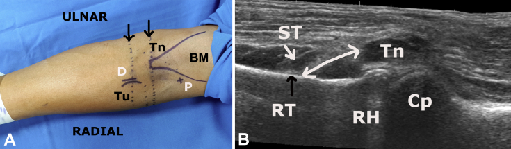
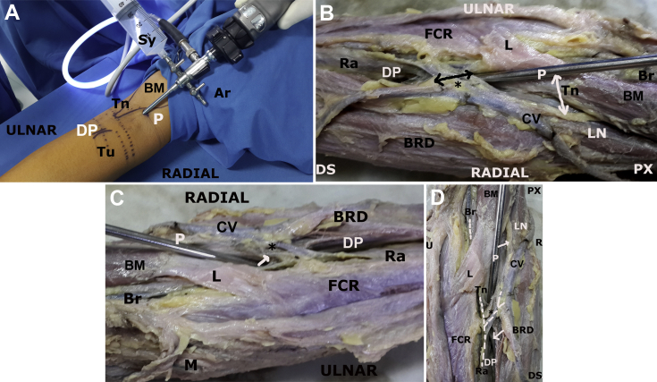
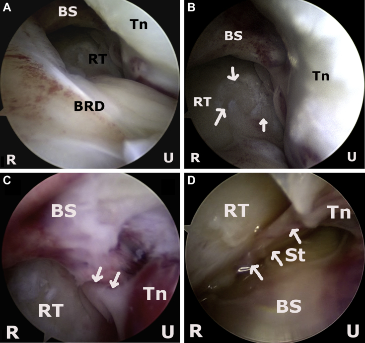
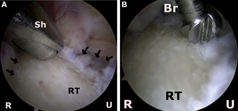
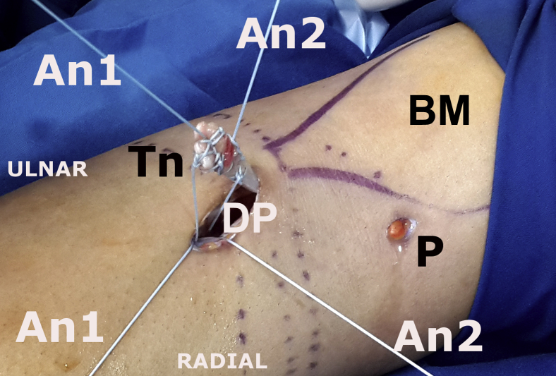
Distal biceps rupture is associated with significant functional disability, and surgical treatment involves open or endoscopic-assisted repair of the ruptured tendon through an anterior incision. This report describes an endoscopic approach that is performed with 2 portals for visualization and instrumentation. Preoperative sonography is used to identify bony and soft-tissue landmarks. The viewing portal is a proximal anterolateral "parabiceps portal" developed by the author, and the landmarks and relevant anatomic relations have been derived from a preliminary anatomic study. The working portal is a distal anterior portal and permits access to the radial tuberosity through the internervous muscular plane. The parabiceps portal permits visualization of the anterior and medial region of the radial tuberosity. A detailed description of the endoscopic pathoanatomy of the distal biceps tendon region is presented. The distal anterior portal is used for retrieval of the ruptured tendon, and thereafter the tuberosity is debrided and anchors are placed under vision. The ruptured tendon is whipstitched and docked onto the tuberosity, and nonsliding knots are used to securely reattach the tendon to bone. Overall, the 2-portal technique provides a method for tendon repair under direct visualization and is safe and reproducible.


Figures

Fig 1
Fig 1
(A) Overview of elbow position (left elbow) and external anatomic landmarks. The transverse level of the radial tuberosity (Tu) and of the ruptured biceps tendon is predetermined sonographically and marked on the forearm (arrows). The parabiceps portal (P) is marked 5 to 7 cm proximal to the radial tuberosity, on the lateral aspect of the biceps muscle (BM) and adjacent to the distal biceps tendon (Tn). The distal anterior portal (D) is placed linearly along the one-third to two-thirds junction of the tuberosity level. (B) Sonographic evaluation of distal biceps region of left elbow. The ruptured distal biceps tendon is seen retracted 1.5 to 2 cm proximal to the tuberosity (double-headed arrow). The biceps sheath or pseudotendon (ST, white arrow) is seen attaching to the radial tuberosity (RT, black arrow). These sonographic landmarks are used to mark the skin levels of the tuberosity and tendon before surgery. (Cp, capitellum; RH, radial head.)
Fig 2
Fig 2
(A) The parabiceps portal (P) is created by passing the arthroscopic sheath in a distal direction toward the tuberosity (Tu) and is angled approximately 20° inferomedially. The sheath is parallel to the distal biceps tendon (Tn), and the passage should be smooth without any resistance. A 50-mL syringe (Sy) is used to insufflate the space with air, and a diagnostic endoscopy is performed. (B) Anatomic relations of the parabiceps portal (P) are shown in a left cadaveric elbow as viewed from the radial aspect. At the entry point (P), the arthroscopic sheath is above and lateral to the distal biceps (BM) and tendon (Tn). The lateral cutaneous nerve (LN) is seen exiting under the biceps muscle (BM) and is 5 to 10 mm away from the portal site (white arrow). One should note the inferior angulation of the arthroscope as it courses parallel to the distal biceps tendon (Tn); this angulation places the sheath below the radial and recurrent radial arteries (Ra, black arrow). The arteriovenous arcade (black arrow, asterisk) is in close approximation to the arthroscope sheath. The distal anterior portal (D) is placed in the internervous plane between the flexor carpi radialis muscle (FCR) and brachioradialis muscle (BRD). (C) Anatomic relations of the parabiceps portal (P) are shown in a left cadaveric elbow as viewed from the ulnar aspect. At the entry point (P), the arthroscopic sheath is above and lateral to the distal biceps muscle (BM) and tendon. One should note the inferior angulation of the arthroscope as it courses below the radial and recurrent radial arteries (Ra, asterisk). The arteriovenous arcade (white arrow, asterisk) is visualized from this aspect and is in close approximation to the arthroscope sheath. (D) Anatomic relations of the parabiceps portal (P) are shown in a left cadaveric elbow as viewed in the coronal plane. At the entry point (P), the arthroscopic sheath is above and lateral to the distal biceps muscle (BM) and tendon. One should note the medial angulation of the arthroscope as it courses below the radial and recurrent radial arteries (Ra, asterisk). The arteriovenous arcade (white arrow, asterisk) is visualized from this aspect and is in close approximation to the arthroscope sheath. The distal biceps tendon is seen attaching (white arrow) to the radial tuberosity through the distal anterior portal (D). (Ar, arthroscope; Br, brachial vessels; BRD, brachioradialis muscle; BM, biceps muscle; CV, cephalic vein; DP, distal portal; DS, distal; FCR, flexor carpi radialis muscle; L, lacertus fibrosus; LN, lateral cutaneous nerve; M, medial epicondyle; PX, proximal; R, radial; U, ulnar.)
Fig 3
Fig 3
Diagnostic tendoscopy of left distal biceps tendon region. (A) The arthroscope is placed in the parabiceps portal and is positioned halfway along the portal tract. The distal biceps tendon (Tn) is seen along the ulnar (U) aspect, and the brachioradialis muscle (BRD) is seen on the radial (R) aspect. The radial tuberosity (RT) is seen in the depth of the field and is under the soft-tissue sleeve of the biceps tendon (BS). (B) The arthroscope is placed in the parabiceps portal and is positioned adjacent to the radial tuberosity. The avulsed footprint (arrows) of the distal biceps on the tuberosity is visualized. The biceps sleeve forms the arched roof of the space. The ulnar-most aspect of the tuberosity and the distal-most tip of the biceps are not visualized from this position. (C) The arthroscope is placed in the parabiceps portal and is positioned ulnar to the radial tuberosity. The bulbous distal end of the biceps is visualized, and the narrow remnant fibers (arrows) are seen extending to the posteromedial aspect of the tuberosity. (D) The arthroscope is placed in the parabiceps portal and is positioned ulnar to the radial tuberosity to visualize the posteromedial aspect of the radial tuberosity. The attachment of any remnant or pseudotendon (ST, arrows) of the distal biceps is visualized and confirmed with pronation-supination.
Fig 4
Fig 4
Overview of the 2 portals used for distal biceps repair. The distal anterior portal (DP) is created using a 6- to 8-mm skin incision adjacent to the sonographically marked tuberosity (Tu). Blunt dissection is performed down to the radial tuberosity, and a short 6- to 8-mm cannula (Cn) is placed through this portal. The parabiceps portal (P) is the viewing portal and the distal anterior portal is the working portal for the entire procedure. (Ar, arthroscope; BM, biceps muscle; Tn, distal biceps tendon.)
Fig 5
Fig 5
(A) Retrieval of left distal biceps tendon region. The arthroscope is placed in the parabiceps portal and is positioned ulnar to the distal biceps (Tn). The biceps tendon is identified as a bulbous structure on the ulnar aspect of the view. A grasper (Gr) is introduced through the distal portal, and the tendon is grasped. (B) The tendon (Tn) is retrieved through the distal anterior portal (DP) and is held with a looped suture to prevent retraction. One should note that the tendon end is frayed and thin and should be excised before repair. (Ar, arthroscope; BM, biceps muscle; P, parabiceps portal; R, radial; U, ulnar.)
Fig 6
Fig 6
(A) Tuberosity (RT) preparation (left elbow, viewed through parabiceps portal) is initiated with an arthroscopic shaver (Sh) placed through the distal anterior portal. A large amount of the tuberosity surface is debrided, and the radial (R) and ulnar (U) limits are identified (arrows) by forearm rotation. (B) Tuberosity (RT) preparation (left elbow, viewed through parabiceps portal) is continued, and decortication of the radial tuberosity is performed with an arthroscopic burr (Br). The hood of the instrument is used to protect the ulnar and superior soft tissues.
Fig 7
Fig 7
(A) Anchor placement (left elbow, viewed through parabiceps portal). The distal anchor (An1) is placed first in the distal region of the radial tuberosity (RT) by use of an anchor sleeve (SL). The placement should be in the ulnar region and approximately beyond the central zone across the breadth of the tuberosity. (B) The distal anchor is seen placed flush with the cortex (arrow) and in the central-ulnar zone of the radial tuberosity. Sutures are retrieved externally through the distal anterior working portal. (C) Anchor placement (left elbow, viewed through parabiceps portal). The proximal anchor (An2) is placed after the distal anchor (An1) in the proximal region of the radial tuberosity (RT) by use of an anchor sleeve (SL). The placement should be in the central-ulnar region. The anchors should be placed at a distance of 10 to 15 mm from each other (arrow). (R, radial; U, ulnar.)
Fig 8
Fig 8
The distal biceps tendon (Tn) is stitched using a locking configuration at 2 levels. One suture from the distal anchor (upper suture of An1) is used to whipstitch the tendon for approximately 10 mm. Similarly, one suture from the proximal anchor (upper suture of An2) is used to whipstitch the tendon for another 5 to 10 mm proximal to the previous whipstitch. The remaining suture from each anchor (lower suture of An1 and lower suture of An2) is used to dock the tendon onto the radial tuberosity. (An1, distal anchor; An2, proximal anchor; BM, biceps muscle; DP, distal anterior portal; P, proximal parabiceps viewing portal.)
Fig 9
Fig 9
(A) The distal biceps tendon (Tn) is seen securely docked onto the prepared radial tuberosity (asterisk). The distal anchor sutures (An1) are tied first using a knot pusher (KP), and a nonsliding distal knot (K1) is placed through the distal anterior portal (left biceps, viewed through parabiceps portal). (B) The distal biceps tendon is seen securely docked onto the radial tuberosity. The proximal anchor sutures are tied after the distal knot (K1) is placed using a knot pusher and a second nonsliding knot (K2) is placed through the distal anterior portal. The arrows show the distance between the 2 anchors, and this length of tendon is now in approximation with the radial tuberosity (left biceps, viewed through parabiceps portal). (C) Final view of repaired distal biceps tendon. The repaired tendon is dynamically assessed by viewing through the distal portal. This portal provides a panoramic view of the tendon-tuberosity construct. The distal (K1) and proximal (K2) nonsliding knots are shown before the remaining sutures are cut (left biceps, viewed through distal anterior portal). (D, distal; P, proximal; R, radial; U, ulnar.)
Fig 10
Fig 10
Biplanar imaging shows the correct positioning of the anchors within the radial tuberosity. The anchors are placed on the ulnar aspect of the tuberosity at a distance of 10 mm from each other. The anchor eyelets are subcortical. (An1, distal anchor; An2, proximal anchor; H, humerus; R, radius; UL, ulna.)

Similar articles

Cited by

References

    1. Baker B.E., Bierwagen D. Rupture of the distal tendon of the biceps brachii. Operative versus non-operative treatment. J Bone Joint Surg Am. 1985;67:414–417. - PubMed
    1. Watson J.N., Moretti V.M., Schwindel L., Hutchinson M.R. Repair techniques for acute distal biceps tendon ruptures: A systematic review. J Bone Joint Surg Am. 2014;96:2086–2090. - PubMed
    1. Phadnis J., Bain G. Endoscopic-assisted distal biceps footprint repair. Tech Hand Up Extrem Surg. 2015;19:55–59. - PubMed
    1. Greenberg J.A., Fernandez J.J., Wang T. EndoButton-assisted repair of distal biceps tendon ruptures. J Shoulder Elbow Surg. 2003;12:484–490. - PubMed
    1. Grégory T., Roure P., Fontès D. Repair of distal biceps tendon rupture using a suture anchor: Description of a new endoscopic procedure. Am J Sports Med. 2009;37:506–511. - PubMed

LinkOut - more resources