

Incidental gastric glomus tumor after laparoscopic sleeve gastrectomy
- PMID: 27284541
- PMCID: PMC4880434
- DOI: 10.4322/acr.2016.028
Incidental gastric glomus tumor after laparoscopic sleeve gastrectomy
Abstract
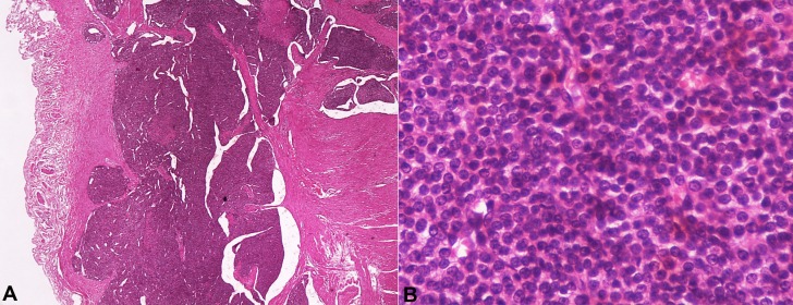
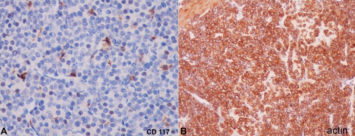
Gastric glomus tumors (GGTs) are unusual benign, subepithelial, mesenchymal neoplasms of modified smooth muscle cells representing a neoplastic counterpart of glomus bodies. A 38-year-old woman was admitted to our clinic presenting morbid obesity. Routine preoperative evaluations, such as laboratory analysis, abdominal ultrasonography, and upper gastrointestinal endoscopy, were performed. She underwent a classical laparoscopic sleeve gastrectomy (LSG). The postoperative course was uneventful and she was discharged for outpatient control. Her histopathology report revealed a GGT 0.8 cm in diameter. No further treatment was done and she had lost 28 kg at the postoperative sixth month. Here, we present the case of GGT, which was diagnosed incidentally after LSG.
Keywords: Bariatric Surgery; Gastrectomy; Glomus Tumor; Incidental Findings; Stomach.
Conflict of interest statement
Conflict of interest: None
Figures
References
-
- Orellana F, Onetto C, Balbontin P, et al. . Gastric glomus tumor: report of one case and review. Endoscopy. 2011;43(Suppl 2):71-2. http://dx.doi.org/10.1055/s-0030-1256041. PMid: - DOI - PubMed
-
- Fang HQ, Yang J, Zhang FF, Cui Y, Han AJ. Clinicopathological features of gastric glomus tumor. World J Gastroenterol. 2010;16(36):4616-20. http://dx.doi.org/10.3748/wjg.v16.i36.4616. PMid: - DOI - PMC - PubMed
-
- Miettinen M, Paal E, Lasota J, Sobin LH. Gastrointestinal glomus tumors: a clinicopathologic, immunohistochemical, and molecular genetic study of 32 cases. Am J Surg Pathol. 2002;26(3):301-11. http://dx.doi.org/10.1097/00000478-200203000-00003. PMid: - DOI - PubMed
-
- Tsuneyoshi M, Enjoji M. Glomus tumor: a clinicopathologic and electron microscopic study. Cancer. 1982;50(8):1601-7. http://dx.doi.org/10.1002/1097-0142(19821015)50:8<1601::AID-CNCR28205008.... PMid: - DOI - PubMed
-
- Kay S, Callahan WP Jr, Murray MR, Randall HT, Stout AP. Glomus tumors of the stomach. Cancer. 1951;4(4):726-36. http://dx.doi.org/10.1002/1097-0142(195107)4:4<726::AID-CNCR2820040410>3.... PMid: - DOI - PubMed
Publication types
LinkOut - more resources
Full Text Sources
Other Literature Sources
Miscellaneous
