

A Comparative Study of Cathepsin D Expression in Peripheral and Central Giant Cell Granuloma of the Jaws by Immunohistochemistry Technique
- PMID: 27284554
- PMCID: PMC4885679
A Comparative Study of Cathepsin D Expression in Peripheral and Central Giant Cell Granuloma of the Jaws by Immunohistochemistry Technique
Abstract
Statement of the problem: Peripheral and central giant cell granuloma are two common benign lesions of the oral cavity. In spite of histopathological similarities, they have different clinical behaviors. Cathepsin D is a lysosomal enzyme which has different functions on the basis of protein and applied peptide cleavage.
Purpose: This research aimed to evaluate and compare the expression level of Cathepsin D in these two lesions to find the reasons for the differences in clinical and biologic characteristics.
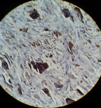
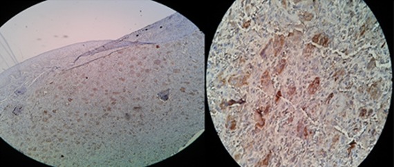
Materials and method: The expression of Cathepsin D was investigated by using the immunohistochemistry method in 20 samples of peripheral giant cell granuloma and 20 samples of central giant cell granuloma. The percentage of stained giant cells (labeling index), the intensity of staining of giant cells, and staining-intensity-distribution in both groups were calculated and compared.
Results: The labeling indices of Cathepsin D in peripheral giant cell granuloma and central giant cell granuloma were 95.9±4.03 and 95.6±2.34, respectively. There was no significant difference in the percentages of stained giant cells between the two groups (p= 0.586). The intensity of staining of giant cells in central giant cell granuloma was stronger than that of peripheral giant cell granuloma (p> 0.001). Staining- intensity- distribution of giant cells in central giant cell granuloma was significantly greater than that of the peripheral type of lesion (p= 0.001).
Conclusion: The higher expression level of Cathepsin D in central giant cell granuloma compared to peripheral type of lesion can explain more aggressive behavior of central giant cell granuloma.
Keywords: Cathepsin D; Giant Cell Granuloma; Immunohistochemistry; Jaw.
Figures
Similar articles
-
A comparative study of osteopontin and MMP-2 protein expression in peripheral and central giant cell granuloma of the jaw.Braz J Otorhinolaryngol. 2019 Mar-Apr;85(2):150-156. doi: 10.1016/j.bjorl.2017.11.006. Epub 2017 Dec 27. Braz J Otorhinolaryngol. 2019. PMID: 29339027 Free PMC article.
-
Are CD68 and Factor VIII-RA Expression Different in Central and Peripheral Giant Cell Granuloma of Jaw: An Immunohistochemical Comparative Study.Turk Patoloji Derg. 2017;1(1):49-56. doi: 10.5146/tjpath.2017.01401. Turk Patoloji Derg. 2017. PMID: 28832079 English.
-
Evaluation of Cyclin D1 Expression in Aggressive and Nonaggressive Central Giant Cell Granuloma of the Jaws.J Dent (Shiraz). 2018 Dec;19(4):253-258. J Dent (Shiraz). 2018. PMID: 30680296 Free PMC article.
-
Hybrid Central Odontogenic Fibroma with Giant Cell Granuloma like Lesion: A Report of Three Additional Cases and Review of the Literature.Head Neck Pathol. 2018 Jun;12(2):166-174. doi: 10.1007/s12105-017-0845-7. Epub 2017 Aug 7. Head Neck Pathol. 2018. PMID: 28785965 Free PMC article. Review.
-
Giant Cell Lesions of the Jaws:A Review and Comparative Histopathological Study.West Afr J Med. 2020 Jan-Mar;37(1):26-31. West Afr J Med. 2020. PMID: 32030708 Review.
Cited by
-
IL-4 induces the formation of multinucleated giant cells and expression of β5 integrin in central giant cell lesion.Med Oral Patol Oral Cir Bucal. 2017 Jan 1;22(1):e1-e6. doi: 10.4317/medoral.20935. Med Oral Patol Oral Cir Bucal. 2017. PMID: 27918730 Free PMC article.
-
Unravelling the role of immunohistochemistry in giant cell lesions of jaws: A systematic review.J Oral Maxillofac Pathol. 2023 Jan-Mar;27(1):181-194. doi: 10.4103/jomfp.jomfp_18_22. Epub 2023 Mar 21. J Oral Maxillofac Pathol. 2023. PMID: 37234327 Free PMC article. Review.
-
Expression of CD34 and CD31 in Central and Peripheral Giant Cell Granulomas.J Dent (Shiraz). 2019 Mar;20(1):10-15. doi: 10.30476/DENTJODS.2019.44557. J Dent (Shiraz). 2019. PMID: 30937331 Free PMC article.
-
A comparative study of osteopontin and MMP-2 protein expression in peripheral and central giant cell granuloma of the jaw.Braz J Otorhinolaryngol. 2019 Mar-Apr;85(2):150-156. doi: 10.1016/j.bjorl.2017.11.006. Epub 2017 Dec 27. Braz J Otorhinolaryngol. 2019. PMID: 29339027 Free PMC article.
References
-
- Motamedi MH, Eshghyar N, Jafari SM, Lassemi E, Navi F, Abbas FM, et al. Peripheral and central giant cell granulomas of the jaws: a demographic study. Oral Surg Oral Med Oral Pathol Oral Radiol Endod. 2007; 103: e39–e43. - PubMed
-
- Torabinia N, Razavi SM, Shokrolahi Z. A comparative immunohistochemical evaluation of CD68 and TRAP protein expression in central and peripheral giant cell granulomas of the jaws. J Oral Pathol Med. 2011; 40: 334–337. - PubMed
-
- Amirchaghmaghi M, Falaki F, Mohtasham N, Imanimoghaddam M. Central Giant Cell Granuloma of the Jaws: A Clinical and Radiographic Study in Khorasan. J Appl Scien. 2010; 10: 777–780.
-
- Noleto JW, Marchiori E, Sampaio RK, Iron KL, Collers FB. Radiological and epidemiological aspects of central giant cell granuloma. Radiol Bras. 2007; 40: 167–171.
LinkOut - more resources
Full Text Sources
Research Materials