

Axin-1 Regulates Meiotic Spindle Organization in Mouse Oocytes
- PMID: 27284927
- PMCID: PMC4902301
- DOI: 10.1371/journal.pone.0157197
Axin-1 Regulates Meiotic Spindle Organization in Mouse Oocytes
Abstract
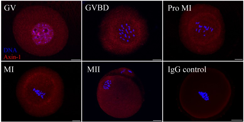
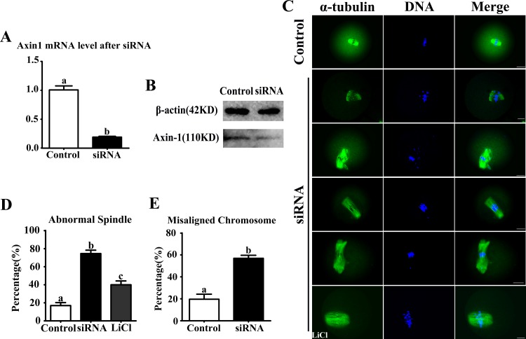
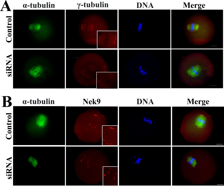
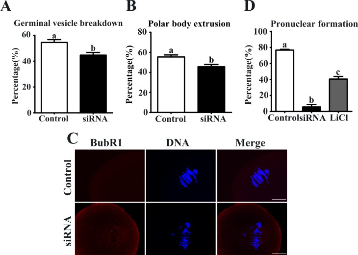
Axin-1, a negative regulator of Wnt signaling, is a versatile scaffold protein involved in centrosome separation and spindle assembly in mitosis, but its function in mammalian oogenesis remains unknown. Here we examined the localization and function of Axin-1 during meiotic maturation in mouse oocytes. Immunofluorescence analysis showed that Axin-1 was localized around the spindle. Knockdown of the Axin1 gene by microinjection of specific short interfering (si)RNA into the oocyte cytoplasm resulted in severely defective spindles, misaligned chromosomes, failure of first polar body (PB1) extrusion, and impaired pronuclear formation. However, supplementing the culture medium with the Wnt pathway activator LiCl improved spindle morphology and pronuclear formation. Downregulation of Axin1 gene expression also impaired the spindle pole localization of γ-tubulin/Nek9 and resulted in retention of the spindle assembly checkpoint protein BubR1 at kinetochores after 8.5 h of culture. Our results suggest that Axin-1 is critical for spindle organization and cell cycle progression during meiotic maturation in mouse oocytes.
Conflict of interest statement
Figures
Similar articles
-
Nek9 regulates spindle organization and cell cycle progression during mouse oocyte meiosis and its location in early embryo mitosis.Cell Cycle. 2012 Dec 1;11(23):4366-77. doi: 10.4161/cc.22690. Epub 2012 Nov 16. Cell Cycle. 2012. PMID: 23159858 Free PMC article.
-
Bora regulates meiotic spindle assembly and cell cycle during mouse oocyte meiosis.Mol Reprod Dev. 2013 Jun;80(6):474-87. doi: 10.1002/mrd.22185. Epub 2013 May 28. Mol Reprod Dev. 2013. PMID: 23610072
-
SUMO-1 plays crucial roles for spindle organization, chromosome congression, and chromosome segregation during mouse oocyte meiotic maturation.Mol Reprod Dev. 2014 Aug;81(8):712-24. doi: 10.1002/mrd.22339. Epub 2014 Jul 30. Mol Reprod Dev. 2014. PMID: 25123474
-
The chromosomal basis of meiotic acentrosomal spindle assembly and function in oocytes.Chromosoma. 2017 Jun;126(3):351-364. doi: 10.1007/s00412-016-0618-1. Epub 2016 Nov 11. Chromosoma. 2017. PMID: 27837282 Free PMC article. Review.
-
Nuclear and spindle positioning during oocyte meiosis.Curr Opin Cell Biol. 2011 Feb;23(1):78-84. doi: 10.1016/j.ceb.2010.07.008. Epub 2010 Aug 11. Curr Opin Cell Biol. 2011. PMID: 20708397 Free PMC article. Review.
Cited by
-
Electrofusion Stimulation Is an Independent Factor of Chromosome Abnormality in Mice Oocytes Reconstructed via Spindle Transfer.Front Endocrinol (Lausanne). 2021 Jul 28;12:705837. doi: 10.3389/fendo.2021.705837. eCollection 2021. Front Endocrinol (Lausanne). 2021. PMID: 34413830 Free PMC article.
-
PATL2 is a key actor of oocyte maturation whose invalidation causes infertility in women and mice.EMBO Mol Med. 2018 May;10(5):e8515. doi: 10.15252/emmm.201708515. EMBO Mol Med. 2018. PMID: 29661911 Free PMC article.
-
Endosomal Wnt signaling proteins control microtubule nucleation in dendrites.PLoS Biol. 2020 Mar 12;18(3):e3000647. doi: 10.1371/journal.pbio.3000647. eCollection 2020 Mar. PLoS Biol. 2020. PMID: 32163403 Free PMC article.
-
Transcription Analysis for Core Networks of lncRNAs-mRNAs: Implication for Potential Role in Sterility of Crassostrea gigas.Biology (Basel). 2022 Feb 27;11(3):378. doi: 10.3390/biology11030378. Biology (Basel). 2022. PMID: 35336752 Free PMC article.
-
MicroRNA-451 is downregulated in the follicular fluid of women with endometriosis and influences mouse and human embryonic potential.Reprod Biol Endocrinol. 2019 Nov 19;17(1):96. doi: 10.1186/s12958-019-0538-z. Reprod Biol Endocrinol. 2019. PMID: 31744497 Free PMC article.
References
-
- Manandhar G, Schatten H, Sutovsky P. Centrosome reduction during gametogenesis and its significance. Biology of reproduction. 2005;72:2–13. - PubMed
MeSH terms
Substances
LinkOut - more resources
Full Text Sources
Other Literature Sources
Molecular Biology Databases
Miscellaneous
