

Development and Implementation of a Corriedale Ovine Brain Atlas for Use in Atlas-Based Segmentation
- PMID: 27285947
- PMCID: PMC4902240
- DOI: 10.1371/journal.pone.0155974
Development and Implementation of a Corriedale Ovine Brain Atlas for Use in Atlas-Based Segmentation
Abstract
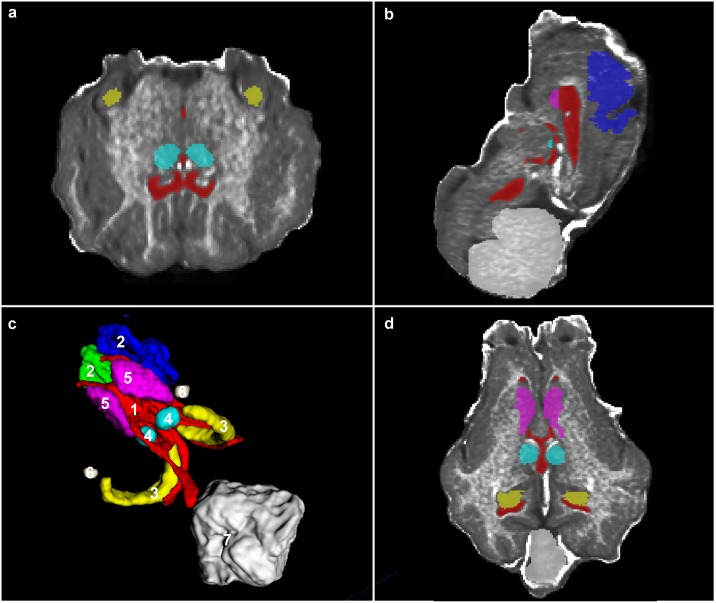
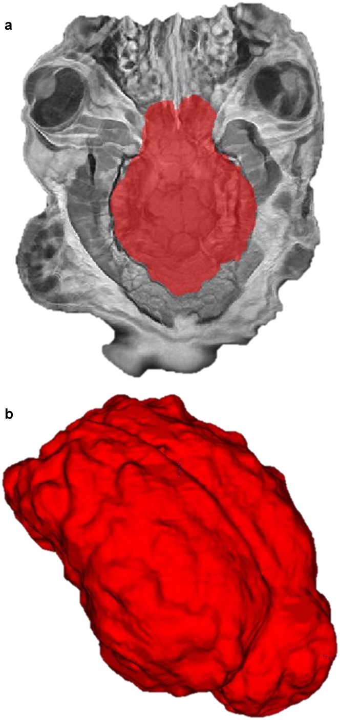
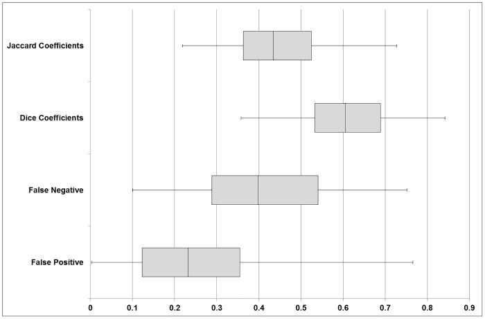
Segmentation is the process of partitioning an image into subdivisions and can be applied to medical images to isolate anatomical or pathological areas for further analysis. This process can be done manually or automated by the use of image processing computer packages. Atlas-based segmentation automates this process by the use of a pre-labelled template and a registration algorithm. We developed an ovine brain atlas that can be used as a model for neurological conditions such as Parkinson's disease and focal epilepsy. 17 female Corriedale ovine brains were imaged in-vivo in a 1.5T (low-resolution) MRI scanner. 13 of the low-resolution images were combined using a template construction algorithm to form a low-resolution template. The template was labelled to form an atlas and tested by comparing manual with atlas-based segmentations against the remaining four low-resolution images. The comparisons were in the form of similarity metrics used in previous segmentation research. Dice Similarity Coefficients were utilised to determine the degree of overlap between eight independent, manual and atlas-based segmentations, with values ranging from 0 (no overlap) to 1 (complete overlap). For 7 of these 8 segmented areas, we achieved a Dice Similarity Coefficient of 0.5-0.8. The amygdala was difficult to segment due to its variable location and similar intensity to surrounding tissues resulting in Dice Coefficients of 0.0-0.2. We developed a low resolution ovine brain atlas with eight clinically relevant areas labelled. This brain atlas performed comparably to prior human atlases described in the literature and to intra-observer error providing an atlas that can be used to guide further research using ovine brains as a model and is hosted online for public access.
Conflict of interest statement
Figures


References
-
- Johnson JI, Rubel EW, Hatton GI (1974) Mechanosensory projections to cerebral cortex of sheep. Journal of Comparative Neurology 158: 81–107. - PubMed
-
- Baskin DS, Browning JL, Widmayer MA, Zhu Z, Grossman RG (1994) Development of a Model for Parkinson's Disease in Sheep using Unilateral Intracarotid Injection of MPTP via Slow Continuous Infusion. Life Sciences 54: 471–479. - PubMed
-
- Opdam HI, Federico P, Jackson GD, Buchanan J, Abbott DF, Fabinyi GC, et al. (2002) A sheep model for the study of focal epilepsy with concurrent intracranial EEG and functional MRI. Epilepsia 43: 779–787. - PubMed
MeSH terms
LinkOut - more resources
Full Text Sources
Other Literature Sources
