. 2016 Oct;94(10):1153-1166.
doi: 10.1007/s00109-016-1439-7. Epub 2016 Jun 10.

Differential transcriptional regulation of hypoxia-inducible factor-1α by arsenite under normoxia and hypoxia: involvement of Nrf2

Affiliations

Differential transcriptional regulation of hypoxia-inducible factor-1α by arsenite under normoxia and hypoxia: involvement of Nrf2

Zukaa Al Taleb et al. J Mol Med (Berl). 2016 Oct.

Abstract

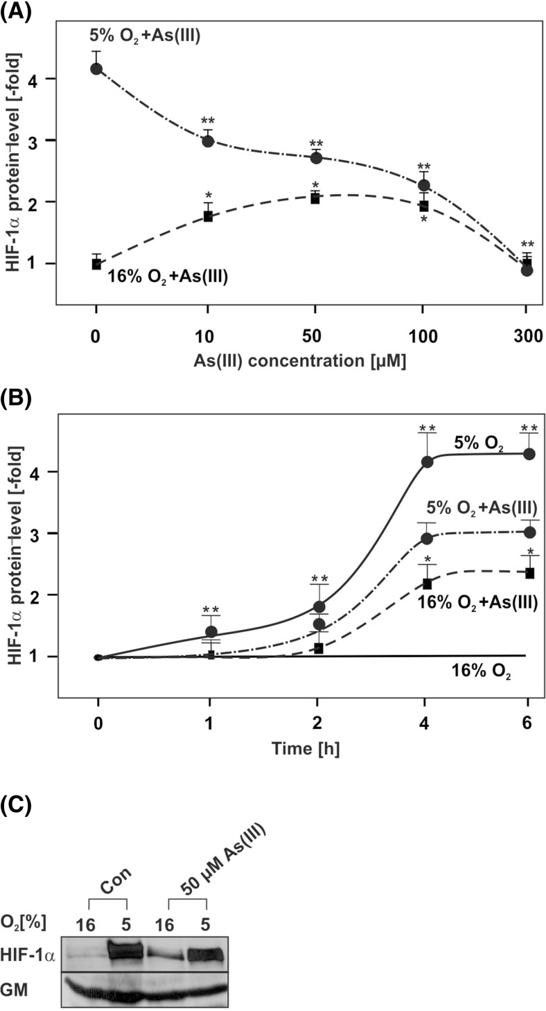
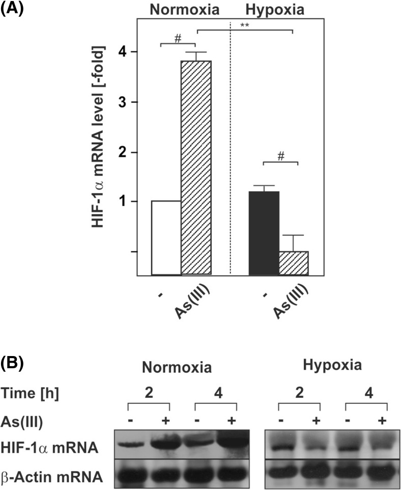
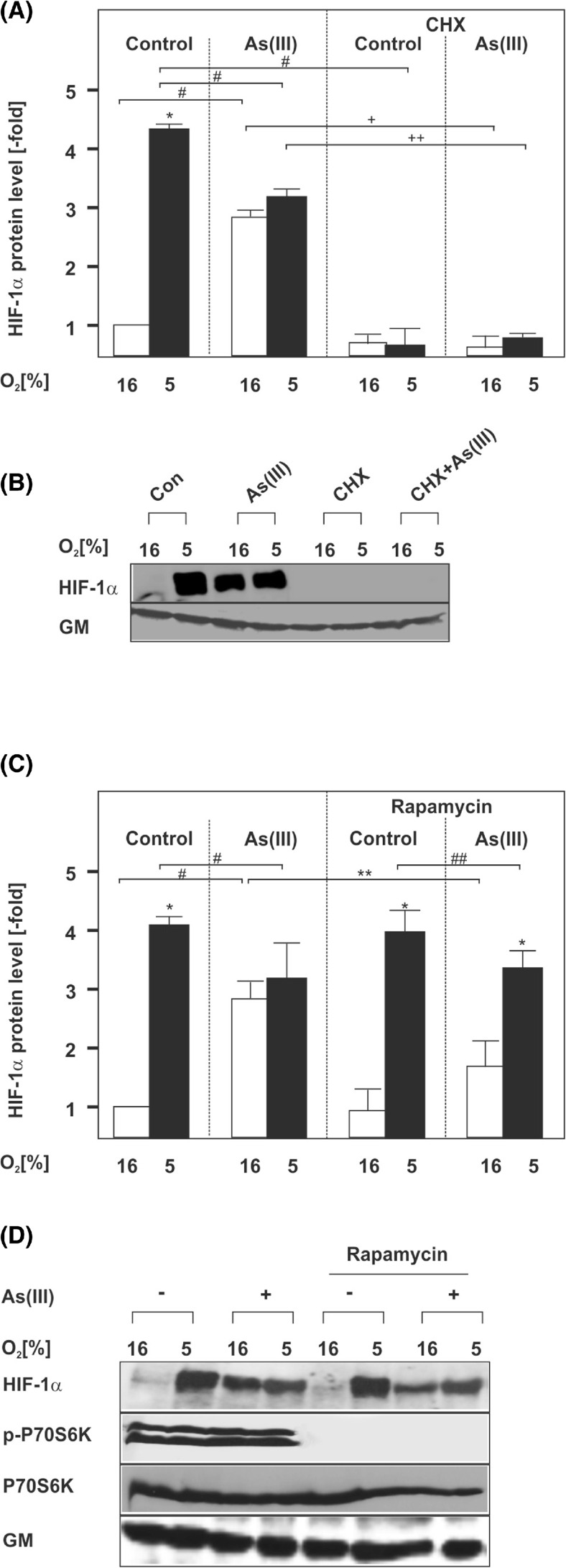
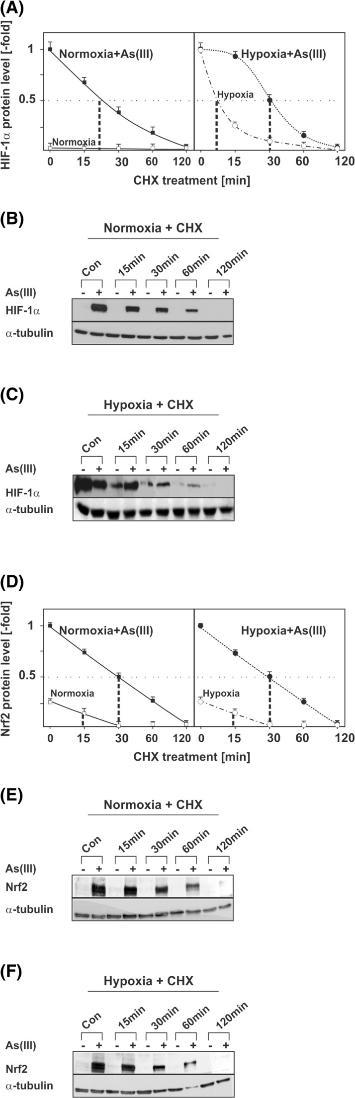
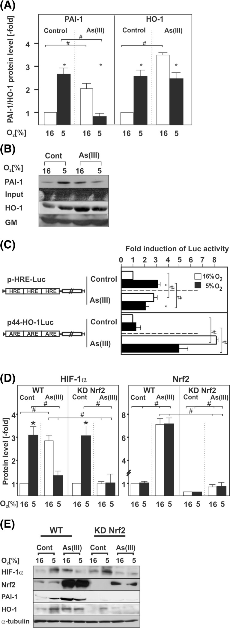
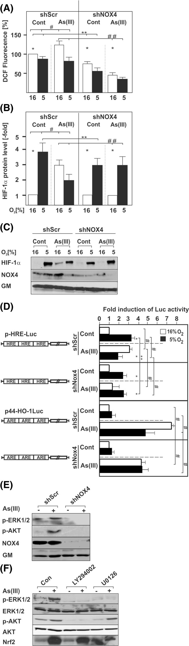
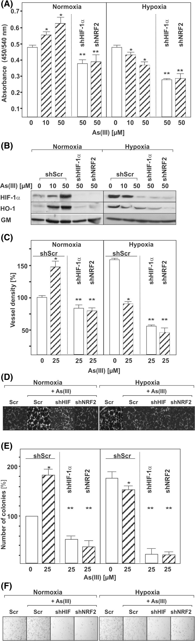
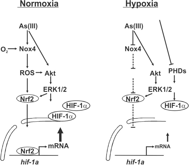
Arsenite (As(III)) is widely distributed in nature and can be found in water, food, and air. There is significant evidence that exposure to As(III) is associated with human cancers originated from liver, lung, skin, bladder, kidney, and prostate. Hypoxia plays a role in tumor growth and aggressiveness; adaptation to it is, at least to a large extent, mediated by hypoxia-inducible factor-1α (HIF-1α). In the current study, we investigated As(III) effects on HIF-1α under normoxia and hypoxia in the hepatoma cell line HepG2. We found that As(III) increased HIF-1α protein levels under normoxia while the hypoxia-mediated induction of HIF1α was reduced. Thereby, the As(III) effects on HIF-1α were dependent on both, transcriptional regulation via the transcription factor Nrf2 mediated by NOX4, PI3K/Akt, and ERK1/2 as well as by modulation of HIF-1α protein stability. In line, the different effects of As(III) via participation of HIF-1α and Nrf2 were also seen in tube formation assays with endothelial cells where knockdown of Nrf2 and HIF-1α abolished As(III) effects. Overall, the present study shows that As(III) is a potent inducer of HIF-1α under normoxia but not under hypoxia which may explain, in part, its carcinogenic as well as anti-carcinogenic actions.

Key message: As(III) increased HIF-1α under normoxia but reduced its hypoxia-dependent induction. The As(III) effects on HIF-1α were dependent on ROS, NOX4, PI3K/Akt, and ERK1/2. The As(III) effects under normoxia involved transcriptional regulation via Nrf2. Knockdown of Nrf2 and HIF-1α abolished As(III) effects in tube formation assays. The data may partially explain As(III)'s carcinogenic and anti-carcinogenic actions.

Keywords: Arsenite As(III); Hypoxia-inducible factor 1 (HIF-1α); Mitogen-activated protein kinase (MAPK); NADPH enzyme oxidase 4 (NOX4); Reactive oxygen species (ROS).


Conflict of interest statement

The authors declare that they have no conflict of interest.

Figures

Fig. 1
Fig. 1
Concentration- and time-dependent modulation of HIF-1α protein levels by As(III) under normoxia and hypoxia. Cells were cultured under normoxia (16 % O2) for 24 h. After 24 h, the medium was changed and cells were further cultured under normoxia and hypoxia (5 % O2) and treated either with: a different concentrations of As(III) for 4 h; b 50 μM As(III) for the indicated time points. The HIF-1α levels in the absence of As(III) or in controls at 0 h were set to 1. Values represent mean ± SEM of at least three independent experiments. Single asterisk, significant difference control under normoxia vs. As(III); double asterisks, significant difference control under hypoxia vs. As(III). c Representative Western blot with 50 μM As(III) at 4 h. One hundred micrograms of total protein was subjected to Western blot analysis with an antibody against HIF-1α or Golgi membrane (GM). Autoradiographic signals were visualized by chemiluminescence and quantified by video densitometry
Fig 2
Fig 2
As(III) induces HIF-1α at the transcriptional level. Cells were cultured under normoxia for 24 h. After 24 h, the medium was changed and cells were further cultured under normoxia and hypoxia in the absence or presence of 50 μM As(III). a, b The HIF-1α mRNA expression levels under normoxia (16 % O2) were set to 1. Values represent means ± SEM of three independent experiments. Single number sign, significant difference As(III) vs. respective control; double asterisks, significant difference As(III) at 16 % O2 vs. As(III) at 5 % O2; p ≤ 0.05. b Representative Northern blot. Twenty micrograms of total RNA from cultured HepG2 cells were subjected to Northern blot analysis and hybridized with DIG-labeled HIF-1α and β-actin probes. Autoradiographic signals were visualized by chemiluminescence and quantified by video densitometry
Fig. 3
Fig. 3
Induction of HIF-1α by As(III) is dependent on mTORC1-driven protein synthesis. Cells were cultured under normoxia for 24 h. At 24 h, the medium was changed and the cells were pre-treated for 30 min either with cycloheximide (CHX, 10 μg/ml) (a, b) or rapamycin (10 μM) (c, d) and then treated with 50 μM As(III) under normoxia (16 % O2) or hypoxia (5 % O2) for 4 h. a, c Statistical analyses of HIF-1α levels. The HIF-1α levels in the controls were set to 1. Values represent mean ± SEM of three independent experiments. Single asterisk, significant difference 16 % O2 vs. 5 % O2; single number sign, significant difference As(III) vs. control; double asterisks, significant difference 16 % O2 + As(III) vs. rapamycin at 16 % O2 + As(III); double number sign, significant difference rapamycin at 5 % O2 vs. rapamycin at 5 % O2 + As(III); single plus sign, significant difference As(III) 16 % O2 vs. As(III)/CHX 16 % O2; double plus sign, significant difference As(III) 5 % O2 vs. As(III)/CHX 5 % O2; p ≤ 0.05. b, d Representative Western blot. One hundred micrograms of isolated total protein was subjected to Western blot analysis with an antibody against HIF-1α, Golgi membrane (GM), phospho-p70S6K1, or total p70S6K1. Signals were visualized by chemiluminescence and quantified by video densitometry
Fig. 4
Fig. 4
Modulation of HIF-1α and Nrf2 half-life by As(III). HepG2 cells were cultured under normoxia for 24 h. At 24 h, the medium was changed and the cells were treated with 50 μM As(III) under normoxia (16 % O2) or hypoxia (5 % O2) for 4 h. After 4 h, cells were treated with cycloheximide (CHX, 10 μg/ml) for the indicated times. a The HIF-1α levels in the As(III)-treated controls were set to 1. Values represent mean ± SEM of three independent experiments. b, c Representative Western Blots. One hundred micrograms of isolated total protein was subjected to Western blot analysis with an antibody against HIF-1α or α-tubulin. d The Nrf2 levels in the As(III)-treated controls were set to 1. Values represent mean ± SEM of three independent experiments. e, f Representative Western Blots. One hundred micrograms of isolated total protein was subjected to Western blot analysis with an antibody against Nrf2 or α-tubulin
Fig. 5
Fig. 5
As(III) affects HIF-target gene expression and knockdown of Nrf2 abolishes As(III)-dependent HIF-1α induction. HepG2 cells were cultured under normoxia for 24 h. At 24 h, the cells were treated with 50 μM As(III) and further cultured for the next 12 h under normoxia (16 % O2) or hypoxia (5 % O2). a Statistical analyses of the PAI-1 and HO-1 protein levels. The PAI-1 and HO-1 levels under normoxia were set to 1. Values are means ± SEM of at least four independent experiments. Single asterisk, significant difference 16 % O2 vs. 5 % O2; single number sign, significant difference As(III) vs. control; p ≤ 0.05. b Representative Western Blot. One hundred micrograms of protein from the cell culture medium or total protein lysates was analyzed by Western blot analysis with an antibody against PAI-1, HO-1, and Golgi membrane (GM). c Cells were transfected with Luc gene constructs containing either three copies of the hypoxia-inducible factor (HIF) binding HRE element (pHRE-Luc) or the Nrf2 binding antioxidant response element (ARE) from the HO-1 gene in front of a minimal promoter (p44-HO-1Luc). The transfected cells were treated with 50 μM As(III) and further cultured for 24 h under normoxia or hypoxia. In each experiment, the LUC activity of the respective control at 16 % O2 was set to 1. Values are means ± SEM of three independent culture experiments; single asterisk, significant difference 16 % O2 vs. 5 % O2; single number sign, significant difference As(III) vs. control. d Statistical analyses of the HIF-1α and Nrf2 protein levels. Cells were cultured under normoxia for 24 h. At 24 h, the cells were treated with 50 μM As(III) and further cultured for the next 8 h under normoxia (16 % O2) or hypoxia (5 % O2). The expression of HIF-1α under normoxia was set to 1. Values are means ± SEM of at least four independent experiments. Single asterisk, significant difference control 16 % O2 vs. 5 % O2; single number sign, significant difference As(III) vs. control. e Representative Western Blots. One hundred micrograms of total protein was analyzed by Western blot with an antibody against HIF-1α, Nrf2, PAI-1, HO-1, and α-tubulin
Fig. 6
Fig. 6
Involvement of ROS, NOX4, PI3K, and ERK1/2 in the response to As(III). Cells transfected with scrambled shRNA (shScr) or shRNAs against NOX4 (shNOX4) were cultured under normoxia for 24 h. At 24 h, the medium was changed and the cells were treated with 50 μM As(III) for 15 min under normoxia (16 % O2) or hypoxia (5 % O2). ROS formation was measured with the DCFH assay. a Statistical analyses of ROS formation. Values represent mean ± SEM of at least three independent experiments. Single asterisk, significant difference shScr vs. shNOX4; double asterisks, significant difference shScr vs. shScr + As(III) or shScr + As(III) vs. shNOX4 + As(III). b Cells transfected as above were treated with 50 μM As(III) and further cultured for 4 h under normoxia (16 % O2) or hypoxia (5 % O2). The HIF-1α protein levels in the controls under normoxia were set to 1. Values represent mean ± SEM of at least three independent experiments. Single asterisk, significant difference 16 % O2 vs. 5 % O2; double asterisks, shScr 5 % O2 vs. shNOX4 5 % O2; double number sign, shScr + As(III) vs. shNOX4 + As(III). c Representative Western blots. One hundred micrograms of total protein was subjected to Western blot analysis with an antibody against HIF-1α, NOX4, or Golgi membrane (GM). d Cells were transfected with scrambled shRNA (shScr) or shRNAs against NOX4 (shNOX4) and Luc gene constructs containing either three copies of the hypoxia-inducible factor (HIF) binding HRE element (pHRE-Luc) or the Nrf2 binding antioxidant response element (ARE) from the HO-1 gene in front of a minimal promoter (p44-HO-1Luc). The transfected cells were treated with 50 μM As(III) and further cultured for 24 h under normoxia or hypoxia. In each experiment, the LUC activity of the respective control at 16 % O2 was set to 1. Values are means ± SEM of three independent culture experiments; single asterisk, significant difference 16 % O2 vs. 5 % O2; double asterisks, shScr 5 % O2 vs. shNOX4 5 % O2; single number sign, shScr vs. shScr + As(III) or shNOX4 + As(III). e Cells transfected with scrambled shRNA (shScr) or shRNAs against NOX4 (shNOX4) were cultured under normoxia for 24 h. At 24 h, the medium was changed and the cells were treated with 50 μM As(III) for 15 min. Thereafter proteins were harvested and subjected to Western analysis with antibodies against phospho-ERK1/2, phospho-Akt, NOX1, NOX4, and Golgi membrane (GM). f Cells were cultured as in e; but after 24 h, they were pre-treated for 30 min with 20 μM of the PI3K inhibitor LY294002 or the MEK inhibitor U0126, before treatment with 50 μM As(III). Proteins were harvested after 15 min for the respective blots. One hundred micrograms of total protein was subjected to Western blot analysis with antibodies against phospho-ERK1/2, phospho-Akt, Nrf2, and Golgi membrane (GM)
Fig. 7
Fig. 7
Influence of As(III) on cell proliferation. a HepG2 cells were cultured in 96-well plates under normoxia for 24 h. At 24 h, the medium was changed and the cells were transfected with plasmids expressing control scrambled shRNA (shCtr) or shRNA against HIF-1α or Nrf2. The next day, cells were cultured in the absence or presence of As(III) under normoxia (16 % O2) or hypoxia (5 % O2). After 4 h, BrdU assay was performed. Values represent mean ± SEM of at least three independent experiments. Single asterisk, significant difference control vs. As(III). Double asterisks, significant difference As(III) vs. As(III) + shNrf2 or As(III) + shHIF-1α. b Representative Western Blot. One hundred micrograms of total protein was subjected to Western blot analysis and probed with an antibody against HIF-1α, HO-1, or Golgi membrane (GM). c HMEC-1 cells were transfected with vectors for control scrambled shRNA (shScr) or shRNA against HIF-1α or Nrf2. Cells were plated onto matrigel-coated wells for 2 h and then exposed to As(III) under normoxia or hypoxia for 4 h. The number of tubules from each well was counted using ImageJ software. Data represent the number of tubules relative to the control which was set to 100 %. Single asterisk, significant difference shScr vs. As(III) + shScr. Double asterisks, significant difference As(III) + shScr vs. As(III) + shNrf2 or As(III) + shHIF-1α. d Photographs from a representative in vitro angiogenesis experiment. e Hep3B liver carcinoma cells were transfected as above. Cells were seeded onto six-well plates, exposed to As(III), and grown for 4 days. Data are presented as sum of colony volume (Σ(V)) and sum of area (Σ(A)), respectively, relative to the control which was set to 100 %. Single asterisk, significant difference shScr vs. As(III) + shScr. Double asterisks, significant difference As(III) + shScr vs. As(III) + shNrf2 or As(III) + shHIF-1α. f Photographs from a representative colony formation assay
Fig. 8
Fig. 8
Scheme of the differential transcriptional regulation of hif-1α by arsenite under normoxia and hypoxia: role of Nrf2. When O2 is not a limiting factor, i.e., under normoxia, As(III) induces HIF-1α mRNA transcription, HIF-1α protein synthesis, HIF-1α target gene expression as well as cell proliferation and angiogenesis by involving ROS formation via NOX4 and the redox-sensitive transcription factor Nrf2. Thereby, NOX4 regulated activation of PI3K/Akt and ERK1/2 appear also to contribute to the stimulating effect. Under O2 limiting conditions, i.e., hypoxia, less ROS can be formed via NOX4 with the consequence that HIF-1α can no longer be efficiently transcribed due to diminished Nrf2 activity. Although the cells try to compensate against this inhibitory effect on HIF-1α transcription by prolonging its protein half-life, cell proliferation and angiogenesis are strongly impaired

Similar articles

Cited by

References

    1. The World Bank . Towards a more effective operational response. Washington, DC: The World Bank; 2005.
    1. Tseng CH, Chong CK, Tseng CP, Hsueh YM, Chiou HY, Tseng CC, Chen CJ. Long-term arsenic exposure and ischemic heart disease in arseniasis-hyperendemic villages in Taiwan. Toxicol Lett. 2003;137:15–21. doi: 10.1016/S0378-4274(02)00377-6. - DOI - PubMed
    1. Feldman RG, Niles CA, Kelly-Hayes M, Sax DS, Dixon WJ, Thompson DJ, Landau E. Peripheral neuropathy in arsenic smelter workers. Neurology. 1979;29:939–944. doi: 10.1212/WNL.29.7.939. - DOI - PubMed
    1. Wasserman GA, Liu X, Parvez F, Ahsan H, Factor-Litvak P, van Geen A, Slavkovich V, LoIacono NJ, Cheng Z, Hussain I, et al. Water arsenic exposure and children’s intellectual function in Araihazar, Bangladesh. Environ Health Perspect. 2004;112:1329–1333. doi: 10.1289/ehp.6964. - DOI - PMC - PubMed
    1. Arsenic and arsenic compounds (1980). IARC Monogr Eval Carcinog Risk Chem Hum 23:39-141 - PubMed

Publication types

MeSH terms

LinkOut - more resources