Review
. 2016 Nov;38(6):635-649.
doi: 10.1007/s00281-016-0574-0. Epub 2016 Jun 10.

The decidua-the maternal bed embracing the embryo-maintains the pregnancy

Affiliations
Review

The decidua-the maternal bed embracing the embryo-maintains the pregnancy

Mayumi Mori et al. Semin Immunopathol. 2016 Nov.

Abstract

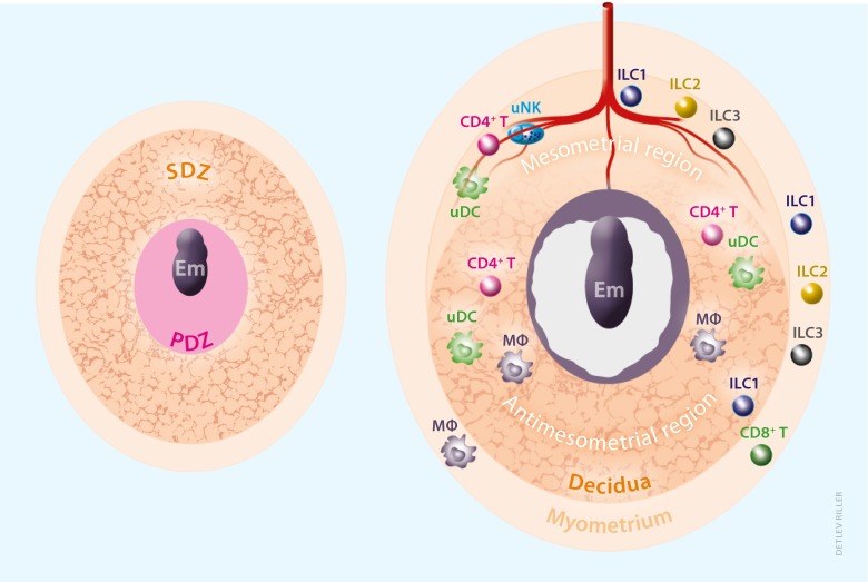
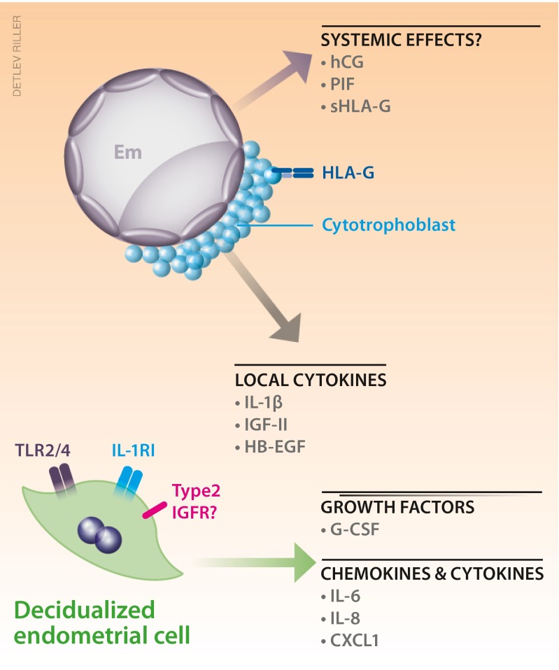
The decidua has been known as maternal uterine tissue, which plays essential roles in protecting the embryo from being attacked by maternal immune cells and provides nutritional support for the developing embryo prior to placenta formation. However, there are questions that still remain to be answered: (1) How does the decidua supply nutrition and provide a physical scaffold for the growing embryo, before placental vascular connection is established? (2) How is the balance between preventing an anti-embryo immune response and protecting both embryo and mother from infections established? To understand basic personas in decidual tissues, we review the structure of the decidua composed of terminally differentiated uterine stromal cells, blood vessels, and a number of repertoire of uterine local immune cells, including the well-known uterine natural killer (uNK) cells and recently discovered innate lymphoid cells (ILCs). Decidual macrophages and uterine dendritic cells (DCs) are supposed to modulate adaptive immunity via balancing cytokines and promoting generation of regulatory T (Treg) cells. During decidualization, vascular and tissue remodeling in the uterus provide nutritional and physical support for the developing embryo. Secretion of various cytokines and chemokines from both the embryo and the decidual cells activates multiple signaling network between the mother and the embryo upon implantation. Defects in the decidual development during early pregnancy result in loss of pregnancy or complications in later gestational stage.

Keywords: Decidua; Endometrium; Infertility; Preeclampsia; Pregnancy; Vascular remodeling.


Figures

Fig. 1
Fig. 1
Anatomical localization of maternal immune cells in murine decidua. Due to the difficulty in obtaining specimens from normal pregnant women at the earliest stage of pregnancy, tissue distribution of maternal immune cells in murine uterus at gestational day (gd) 5.5 and 7.5 is shown. The primary decidual zone (PDZ) is avascular and CD45+ cells are scarcely found [4]. Secondary decidual zone (SDZ) at gd 5.5 and antimesometrial decidua at gd 7.5 are rich in small blood vessels, whereas mesometrial region at gd 7.5 is surrounded by lateral dilated large vessels (not shown). In the mesometrial decidua, uNK cells are most abundantly found [5], also see Fig. 2], DCs are confined in the entire decidua [6]. In addition to uILC1 in the decidua, other uILCs are detected in mesometrial region and myometrium [7], but the interaction of uILCs with vasculature or other immune cells have not been reported. Except for the report on suppressed infiltration of cytotoxic effector T cells in the decidua [8], T cell subsets inside the murine decidua have not been well known. Macrophages can be found in the region between trophoblast and uterine stromal cells in association with vascular endothelial cells [4]. Note that the numbers of representative cell types are not consistent with the actual populations
Fig. 2
Fig. 2
Distribution of uNK cells at early gestational stage in mice. Uterine sections from C57BL/6 female mice at gd 5.5 (a) and gd 6.5 (bd) were stained with DBA lectin (A−C) or PAS (D). a At gd 5.5, DBA+ NK cells are scarcely detected. b At gd 6.5, DBA+ NK cells are increasingly detected in the mesometrial region. c Higher magnification of b. d Higher number of PAS+ NK cells are detected in the continuous section of c. Thin arrows: PAS+ cells, thick arrows: DBA+ cells. Bars, a, b 500 μm, c, d 100 μm
Fig. 3
Fig. 3
Possible human decidua-embryo interaction indicated by in vitro observation. Despite of difficulty in obtaining human specimen during normal peri-implantation stage, a number of studies have utilized in vitro co-culture system to investigate signaling communication between the decidual cells and the embryo. Fertilized eggs secrete interleukin-1β (IL-1β) and growth factors such as IGF-II and HB-EGF, which are indicated to regulate decidual cellular development, in line with expression of IL-1RI and type 2 IGFR in the endometrial cells. Signaling for decidual expression of Toll-like receptors (TLRs) is unknown. Soluble factors from the embryo but detected in maternal peripheral blood, such as human chorionic gonadotropin (hCG), preimplantation factor (PIF), and soluble form of HLA-G (sHLA-G), may have systemic effects such as promoting ovarian progesterone production, balancing cytokines and chemokines secretion in the periphery. Decidual cells also express growth factors such as G-CSF, and cytokines and chemokines such as IL-6, IL-8, and CXCL1. At least some of them are considered to be under the control of signaling from the embryos

References

    1. McLaren A, Tarkowski AK. Implantation of mouse eggs in the peritoneal cavity. J Reprod Fertil. 1963;6:385–392. doi: 10.1530/jrf.0.0060385. - DOI - PubMed
    1. Spornitz UM. Pseudo-decidualization at the site of implantation in tubal pregnancy. Arch Gynecol Obstet. 1993;253:85–95. doi: 10.1007/BF02768734. - DOI - PubMed
    1. Deanesly R. Termination of early pregnancy in rats after ovariectomy is due to immediate collapse of the progesterone-dependent decidua. J Reprod Fertil. 1973;35:183–186. doi: 10.1530/jrf.0.0350183. - DOI - PubMed
    1. Douglas NC, Zimmermann RC, Tan QK, Sullivan-Pyke CS, Sauer MV, Kitajewski JK, Shawber CJ. VEGFR-1 blockade disrupts peri-implantation decidual angiogenesis and macrophage recruitment. Vasc Cell. 2014;6:16. doi: 10.1186/2045-824X-6-16. - DOI - PMC - PubMed
    1. Felker AM, Chen Z, Foster WG, Croy BA. Receptors for non-MHC ligands contribute to uterine natural killer cell activation during pregnancy in mice. Placenta. 2013;34:757–764. doi: 10.1016/j.placenta.2013.06.004. - DOI - PMC - PubMed

Publication types

LinkOut - more resources