. 2016 Dec 15;22(24):6099-6109.
doi: 10.1158/1078-0432.CCR-16-0630. Epub 2016 Jun 10.

Dual NAMPT and BTK Targeting Leads to Synergistic Killing of Waldenström Macroglobulinemia Cells Regardless of MYD88 and CXCR4 Somatic Mutation Status

Affiliations

Dual NAMPT and BTK Targeting Leads to Synergistic Killing of Waldenström Macroglobulinemia Cells Regardless of MYD88 and CXCR4 Somatic Mutation Status

Michele Cea et al. Clin Cancer Res. .

Abstract

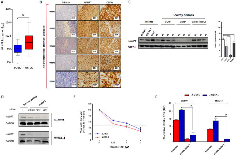
Purpose: Nicotinamide phosphoribosyltransferase (Nampt) regulates intracellular NAD+ pool and is highly expressed in a number of malignancies. FK866, a selective inhibitor of Nampt, depletes intracellular NAD+ levels, thereby blocking cellular metabolism and triggering sensitization to other drugs and cell death. Here we characterized the antitumor effects of Nampt inhibition in Waldenström macroglobulinemia.

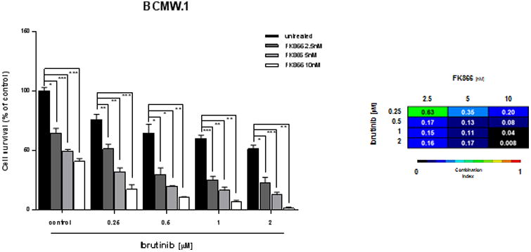
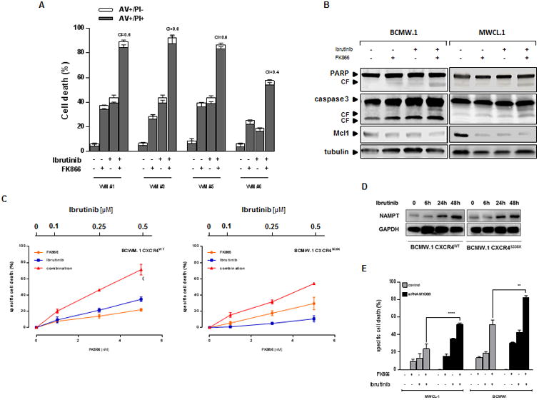
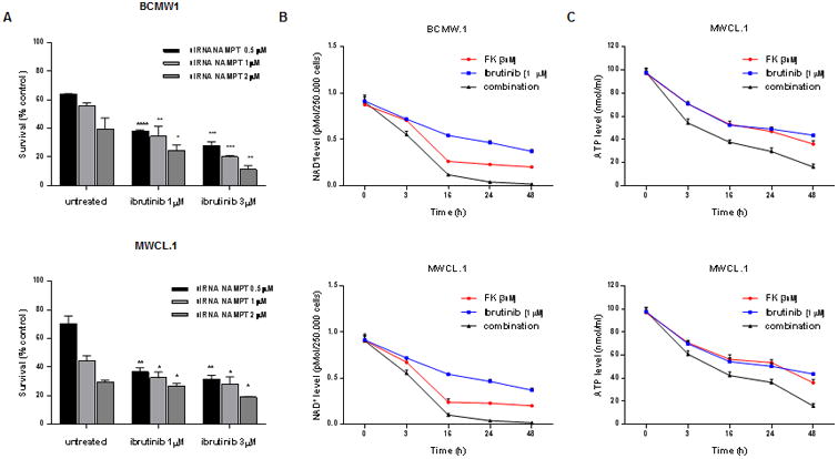
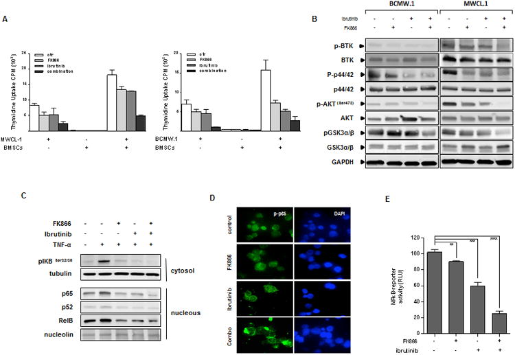
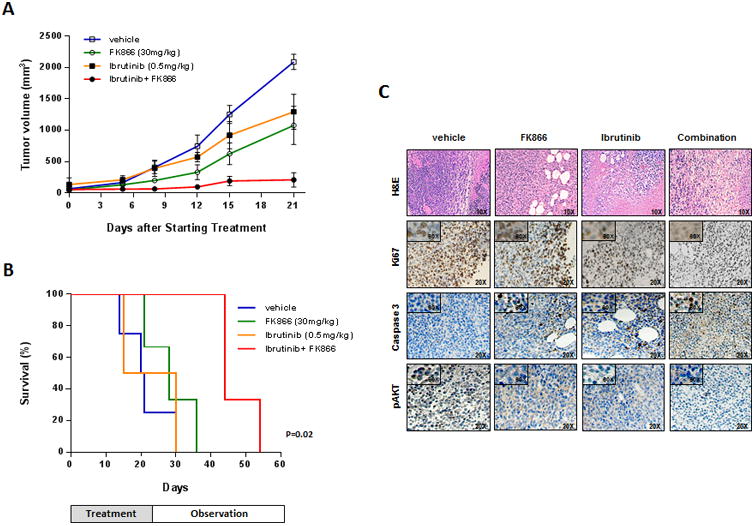
Experimental design: We investigated Nampt role in MW cells using both mRNA and protein expression analyses. We have also used loss-of-function approaches to investigate the growth and survival effects of Nampt on MW cells and further tested the anti-MW activity of dual Nampt and BTK inhibition in vitro and in vivo RESULTS: We found that Waldenström macroglobulinemia cells exhibit high levels of Nampt compared with normal B cells. Loss of function studies suggested a potential oncogenic role of Nampt in Waldenström macroglobulinemia cells, and BTK-inhibitor ibrutinib and FK866 resulted in a significant and synergistic anti-Waldenström macroglobulinemia cell death, regardless of MYD88 and CXCR4 mutational status. Cell death was associated with: (i) activation of caspase-3, PARP and downregulation of Mcl-1, (ii) enhanced intracellular ATP and NAD+ depletion, (iii) inhibition of NF-κB signaling, and (iv) inhibition of multiple prosurvival signaling pathways. In a murine xenograft Waldenström macroglobulinemia model, low-dose combination FK866 and ibrutinib is well tolerated, significantly inhibits tumor growth, and prolongs host survival.

Conclusions: Our results show intracellular NAD+ level as crucial for proliferation and survival of Waldenström macroglobulinemia cells, and provides the mechanistic preclinical rationale for targeting Nampt, either alone or with Ibrutinib, to overcome drug resistance and improve patient outcome in Waldenström macroglobulinemia. Clin Cancer Res; 22(24); 6099-109. ©2016 AACR.


Figures

Figure 1
Figure 1
Nampt is overexpressed in WM cells and its specific knockdown impairs tumor cell growth and viability. (A) Dot plot represents Nampt (probe ID 217739_s_at) expression value from microarray analysis of WM profiles retrieved from NCBI database (GSE9656). The samples were divided into two groups: control (left panel) including normal CD19+ peripheral blood B cells (PB-BC; n=17) and tumor (right panel) including CD19+ WM cells (WM-BC; n=41). (B) Giemesa, CD79α and Nampt IHC analysis (positive cells are brown) on four representative lymph node specimens derived from WM patients (WM 1-4). Magnification: 20× (also 100× for WM 1). (C) Low grade lymphoma cell lines (BCMW.1, MWCL-1, Mec1 and RL), CD19+/-, and PBMCs from 3 healthy donors were subjected to WB analysis (one of three representative blots) using anti-Nampt Ab. Anti-GAPDH monoclonal antibody served as a loading control. Average of Nampt relative expression was calculated as the ratio of the densitometry signal for Nampt relative to GAPDH in each sample using Image J software (1.37v; National Institutes of Health, http://rsb.info.nih.gov/ij/). PBMCs, peripheral blood mononuclear cells; GAPDH, glyceraldehyde 3-phosphate dehydrogenase; HCL, human cell lines. (D) BCMW.1 and MWCL-1 cell lines were transfected using different concentrations of SMARTpool Nampt siRNA or non-targeting siRNA. Two days later, cells were subjected to immunoblotting to assess decrease in Nampt protein expression post-transfection using anti Nampt and GAPDH Abs. WB analyses confirmed reduction in Nampt protein level following transient transfection of WM cells with Nampt siRNA compared with cells transfected with scramble control. (E) The effect of Nampt knockdown on cell survival in WM cells transfected with Nampt or control siRNA were assessed by CellTiter Glo assay and presented as change relative to scramble control cells. (F) WM cell lines transfected with 2 μM of SMARTpool Nampt siRNA or control siRNA were cultured in the absence or presence of BMSCs for 24 hours. Tumor cell growth was evaluated by [3H]thymidine uptake and presented as a percentage of cell growth compared with scramble control. All data in A-F panels are shown as the mean values ± S.D. of triplicates. (one representative experiment performed in triplicate). *p=0.02, **0.001 <p< 0.003, **** p<0.0001 (Student's t test).
Figure 2
Figure 2
Dual inhibition of Nampt and BTK leads to synergistic killing in WM cells. (A) BCMW.1 cells were treated with an inhibitor of Nampt (FK866), BTK (ibrutinib), or both. Cell death was measured by cell viability assay and presented as a percentage of control cells (untreated). Synergism was calculated by CI analysis with heat maps depicting the CI values at increasing doses of FK866 and ibrutinib. Data are presented as mean ± S.D (n=3). CIs were generated with CalcuSyn software for each set of combination. CI<1, =1 and >1 denote synergism, additive effect, and antagonism, respectively. ns, not significant, *0.04
Figure 3
Figure 3
Treatment with FK866 plus ibrutinib induces apoptotic cell death in WM patients derived cells, regardless of MYD88 and CXCR4 somatic mutational status. (A) Ex vivo tumor cells (CD19+ gated) from WM patients were treated with FK866 plus ibrutinib, or either agent alone. Cell death was assessed by flow cytometry using annexin V and PI double staining after 72 hours. The percentage of early apoptotic cells (AV+/PI-) are shown as white columns and that of late apoptotic cells (AV+/PI+) are shown as solid gray columns. Data are derived from at least 3 independent experiments. (P < .05 for all patient samples). A CI less than 1 indicates synergism. (B) BCMW.1 and MWCL-1 cells were treated with either FK866 (3 nM), ibrutinib (1 μM) and the combination for 48 h. Cells were then harvested, and whole cell lysates were subjected to immunoblot analysis using anti-PARP, anti-caspase 3, anti-Mcl-1, or anti-actin antibodies. FL indicates full length and CF cleaved fragment. (C) CXCR4WT and CXCR4S338X expressing WM cells were treated with low dose FK866 (1-5 nM) plus ibrutinib (0.1-0.5 μM) or each drug alone for 72 h. Cell viability was assessed by CellTiter Glo assay and presented as a percentage of control cells (untreated cells). (D) CXCR4WT and CXCR4S338X BCMW.1 cells were treated with ibrutinb (1 μM) for 12 h, 24 h and 48 h. Cells were then harvested, and whole cell lysates were subjected to immunoblot analysis using anti-Nampt antibody. (E) BCMW.1 and MWCL-1 cells were transfected with either scramble control or MYD88-specific siRNA; 24 hours after transfection, cells were treated with either FK866, ibrutinib, or the combination, and then cultured for additional 72 hours. Cell viability was measured with specific assay and presented as a percentage of control. All data presented are a representative study set of 3. ** p=0.0032, ****p<0.0001 (Student's t test).
Figure 4
Figure 4
Nampt and its outputs, NAD+ and ATP intracellular levels, are crucial in enhancing ibrutinib-sensitivity of WM cells. (A) BCMW.1 (up) and MWCL-1 (low) cell lines were transfected using different concentrations of Nampt siRNA or non-targeting siRNA. 48 hours later, cells were treated with increased doses of ibrutinib (1-3 μM). After an additional 48 hours, cell viability was measured using a specific assay. Viability of WM cells Nampt depleted was compared with WM cells transfected with scramble control. (B-C) BCMW.1 and MWCL-1 cell lines were treated with FK866 (3 nM), ibrutinib (1 μM) or combined therapy for 3 h. Cells were then harvested, and intracellular NAD+ (B) and ATP (C) levels were measured using an enzyme cyclic and luminescent assay, respectively. All data presented are mean ± SD of 3 independent experiments. *0.04
Figure 5
Figure 5
Combined treatment decreases WM cell growth in the context of the BM microenvironment via inhibition of pro-survival signaling pathways (A) MWCL-1 and BCMW.1 cells were cultured in the absence (-) or presence (+) of BMSCs from WM patients with FK866, ibrutinib, or the combination. Cell proliferation was assessed by [3H]-thymidine incorporation assay. Data are mean ± SD of triplicate samples. Error bars represent SD. (B) BCMW.1 and MWCL-1 cells were treated with FK866, ibrutinib or combined therapy for 24 h. Whole-cell lysates were then immunoblotted using with the indicated antibodies. Blots shown are representative of 3 independent experiments. (C) Inhibition of NF-kB activity was assessed using western blot analysis of BCMW.1 cells treated with either FK866, ibrutinib, or their combination for 6 h, and then exposed to TNF-α (10 ng/mL) during the last 20 min. Cytoplasmic and nuclear extracts were subjected to Western blotting using specific antibody for analysis of NF-kB canonical (anti–p-NF-κBp65 and -p-IkB) and non-canonical (-NF-κBp52, -RelB) activity. (D) BCMW.1 cells were cultured with FK866, ibrutinib, or the combination for 6 h, with TNF-α (10 ng/mL) added for the last 20 min. Immunocytochemical analysis was performed using anti-pospho-NF-κBp65 antibody. DAPI (4′,6-diamidino-2-phenylindole) was used to stain nuclei. (E) p65-NF-κB activity was also assessed by a luciferase promoter assay at 6 hours in BCMW.1 cells following treatment with FK866 (3 nM), ibrutinib (0.5 μM),, or the combination. Experiments were performed in triplicate. A representative experimental set is shown. **p=0.0038, ***p= 0.002, ****p<0.0001 (Student's t test).
Figure 6
Figure 6
FK866 plus ibrutinib trigger synergistic inhibition of human WM cell growth in vivo. (A) Average and SD of tumor volume (mm3) from groups of mice (n=6/group) versus time (days) when tumor was measured. BCMW.1 cells (5 × 10⁶ in 100 μL of serum free RPMI-1640 medium) were implanted in the flank of CB17-SCID mice. After tumor detection, mice were randomized to IP treatment with vehicle, FK866, ibrutinib, or the combination at the indicated doses for three weeks. A significant decrease in tumor growth was noted in combination-treated mice versus vehicle-treated mice (p=0.002 after the first week and p<0.0001 at the end of treatment). Data are mean tumor volume ± SD. (B) Kaplan-Meier survival plot showing survival for mice treated with vehicle, FK866, ibrutinib or their combination at the indicated concentrations. FK866 plus ibrutinib-treated mice show significantly increased survival (p=0.02) compared with vehicle-treated mice. The mean overall survival (OS) was 20 days in the vehicle- versus 44 days in combination-treated cohorts. Error bars represent mean ± SD. (C) Hematoxylin Eosin (10×) and immunohistochemical staining for Ki-67, caspase 3 and pAKT (20×; 60× in insets) in tumors sectioned on day 30 (endpoint) from vehicle-, FK866- (30 mg/kg), ibrutinib- (0.5 mg/kg) and combination-treated mice. Photographs are representative of 2 mice receiving each treatment.

Similar articles

Cited by

References

    1. Berger F, Ramirez-Hernandez MH, Ziegler M. The new life of a centenarian: signalling functions of NAD(P) Trends in biochemical sciences. 2004;29:111–8. - PubMed
    1. Cairns RA, Harris IS, Mak TW. Regulation of cancer cell metabolism. Nature reviews Cancer. 2011;11:85–95. - PubMed
    1. Chiarugi A, Dolle C, Felici R, Ziegler M. The NAD metabolome--a key determinant of cancer cell biology. Nature reviews Cancer. 2012;12:741–52. - PubMed
    1. Hanahan D, Weinberg RA. Hallmarks of cancer: the next generation. Cell. 2011;144:646–74. - PubMed
    1. Houtkooper RH, Canto C, Wanders RJ, Auwerx J. The secret life of NAD+: an old metabolite controlling new metabolic signaling pathways. Endocrine reviews. 2010;31:194–223. - PMC - PubMed

MeSH terms

Substances