. 2016 Jun 10;13(1):144.
doi: 10.1186/s12974-016-0613-8.

Oncostatin M promotes excitotoxicity by inhibiting glutamate uptake in astrocytes: implications in HIV-associated neurotoxicity

Affiliations

Oncostatin M promotes excitotoxicity by inhibiting glutamate uptake in astrocytes: implications in HIV-associated neurotoxicity

Shamsudheen Moidunny et al. J Neuroinflammation. .

Abstract

Background: Elevated levels of oncostatin M (OSM), an interleukin-6 cytokine family member, have been observed in HIV-1-associated neurocognitive disorders (HAND) and Alzheimer's disease. However, the function of OSM in these disease conditions is unclear. Since deficient glutamate uptake by astrocytes is instrumental in HAND-associated neurotoxicity, we hypothesized that OSM impairs glutamate uptake in astrocytes and thereby promotes neuronal excitotoxicity.

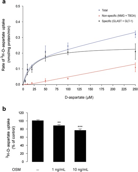
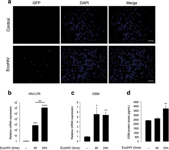
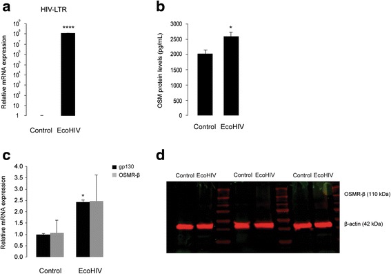
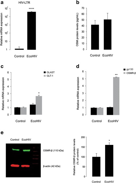
Methods: Primary cultures of mouse cortical astrocytes, neurons, microglia, and BV2 cell line were used. The expression of glutamate transporters (GLAST/EAAT1 and GLT-1/EAAT2) was investigated using real-time PCR and Western blot, and their activity was assessed by measuring (3)H-D-aspartate uptake. Neuronal toxicity was measured using the colorimetric MTT (3-(4,5-dimethylthiazol-2-yl-) 2,5-diphenyltetrazolium bromide) assay and immunocytochemistry. A chimeric HIV-1 that infects murine cells (EcoHIV/NL4-3-GFP virus (EcoHIV)) was used to investigate whether the virus induces OSM, OSM receptor (OSMR)-β, glycoprotein 130 (gp130), GLT-1, GLAST (mRNA and protein), and OSM release (ELISA) in cultured BV2 cells, primary microglia, or astrocytes. Statistical analyses of the data were performed using one-way ANOVA (to allow multiple comparisons) and two-tailed Student's t test.

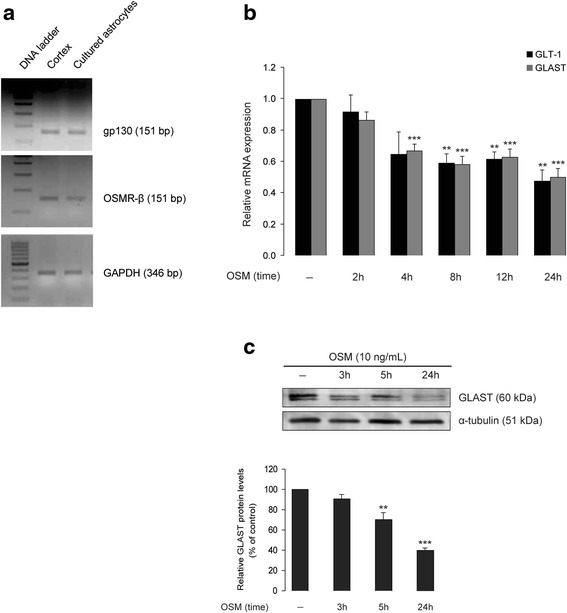
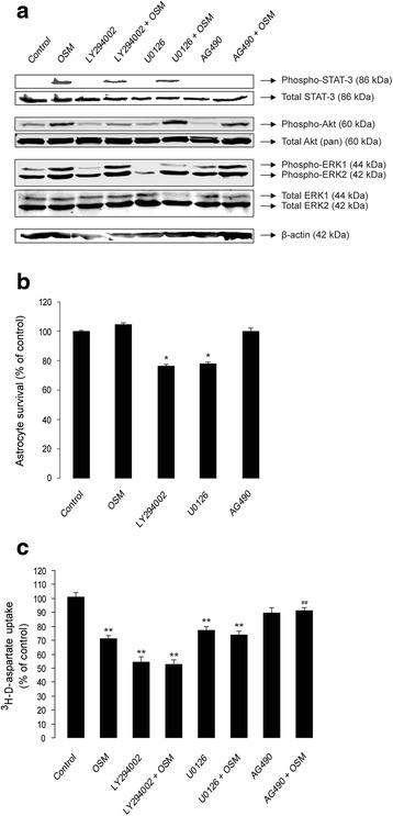
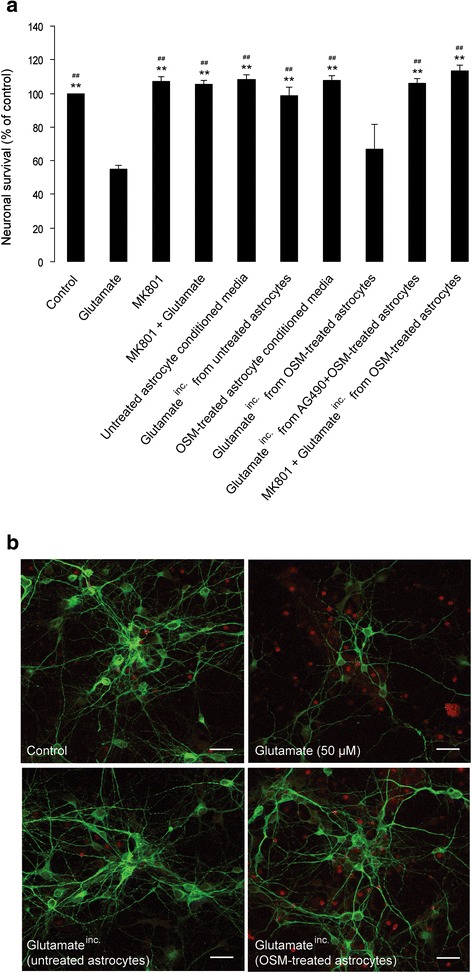
Results: OSM treatment (10 ng/mL) time-dependently reduced GLAST and GLT-1 expression and inhibited (3)H-D-aspartate uptake in cultured astrocytes in a concentration-dependent manner, an effect prevented by the Janus kinase (JAK)/signal transducers and activators of transcription (STAT)3 inhibitor AG490. Down-regulation of astrocytic glutamate transport by OSM resulted in NMDA receptor-dependent excitotoxicity in cortical neurons. Infection with EcoHIV induced OSM gene expression and protein release in BV2 cells and microglia, but not in astrocytes. Conversely, EcoHIV caused a fivefold increase in OSMR-β mRNA (but not gp130) and protein in astrocytes, but not in microglia, which did not express OSMR-β protein. Finally, astrocytic expression of GLAST gene was unaffected by EcoHIV, whereas GLT-1 mRNA was increased by twofold.

Conclusions: We provide first evidence that activation of JAK/STAT3 signaling by OSM inhibits glutamate uptake in astrocytes, which results in neuronal excitotoxicity. Our findings with EcoHIV suggest that targeting OSMR-β signaling in astrocytes might alleviate HIV-1-associated excitotoxicity.

Keywords: Astrocytes; Excitotoxicity; GLAST; GLT-1; Glutamate; HIV; Interleukin 6; NMDA; Oncostatin M.


Figures

Fig. 1
Fig. 1
OSM down-regulates GLT-1 and GLAST expression in primary mouse cortical astrocytes. a Total mRNA purified from wild-type (C57BL/6J) mouse neonatal (P2) brain cortex as well as from cultured cortical astrocytes established from P2 brains was analyzed for expression of OSMR-β and gp130 mRNA by reverse transcriptase PCR; GAPDH primers were used as loading control. b Cortical astrocyte cultures were treated without or with OSM (10 ng/mL) for 2, 4, 8, 12, and 24 h and analyzed for GLT-1 and GLAST mRNA levels (gene expression normalized to HPRT1) by real-time PCR. Data are normalized to untreated controls and presented as mean ± SEM; n = 3, **p < 0.01, ***p < 0.001; one-way ANOVA using Bonferroni correction. c Cortical astrocytic cultures were treated without or with OSM (10 ng/mL) for 3, 5, and 24 h and were analyzed for GLAST proteins by Western blot. Relative densitometric analysis of GLAST proteins is shown in the lower panel. Data are presented as percentage of each respective ratio between optical density value of GLAST band intensity and optical density value of the matched α-tubulin (which served as the loading control) band intensity; n = 3, **p < 0.01, ***p < 0.001; one-way ANOVA
Fig. 2
Fig. 2
OSM inhibits d-aspartate uptake in primary mouse cortical astrocytes. a Shows saturation isotherm of Na+-dependent 3H-d-aspartate uptake by astrocytes. Primary astrocytes from wild-type (C57BL/6J) mouse neonatal (P2) brains were incubated with 3H-d-aspartate (0 to 200 μM) dissolved either in a Na+ buffer or in a Na+-free buffer (where Na+ is replaced by N-methyl-d-glucamine chloride, NMG, to determine nonspecific tritium retention) for 10 min at room temperature. Each point represents the mean ± SEM of at least three separate experiments, measured in triplicate. Kinetic constants were determined by nonlinear regression fit or a rectangular hyperbola, where V max was 0.3 μM ± 0.08 and K M was 22.4 μM (95 % confidence interval: 0.17–0.34 μM). b Shows the effect of OSM treatment (1 and 10 ng/mL; for 24 h) on 3H-d-aspartate uptake in cortical astrocytes culture. Data are normalized to untreated controls and presented as mean ± SEM; *p < 0.05, **p < 0.01, n = 3 to 17; one-way ANOVA followed by Dunnett’s multiple comparison test
Fig. 3
Fig. 3
Inhibition of JAK/STAT3, but not PI3K/Akt or MEK/ERK1/2 signaling pathways, prevents OSM-induced reduction of 3H-d-aspartate uptake in primary cortical astrocytes. a Shows the effect of 2 h pre-treatment with selective inhibitors of PI3K (LY294002, 25 μM), MEK1/2 (U0126, 5 μM), and JAK (AG490, 25 μM) on activation of Akt, ERK1/2, or STAT3 proteins, respectively, in OSM-treated (10 ng/mL for 1 h) astrocytes culture. Activation of different signaling proteins was evaluated by Western blot using antibodies that selectively detect the phosphorylation at Ser473, Thr202/Tyr204, and Tyr705 residues of Akt, Erk1/2, and STAT3, respectively. Total amounts of each protein were detected using appropriate antibodies (see the “Methods” section); β-actin served as loading control. b Shows the effect of OSM (10 ng/mL), LY294002 (25 μM), U0126 (5 μM), and AG490 (25 μM) treatments for 24 h on the viability of cultured cortical astrocytes. Cell viability was measured using a colorimetric MTT assay. OD measurements were made at 570 nm, with a blank correction made at 630 nm. Data are presented as percentage of untreated (control) astrocytes; each bar represents the average of four independent experiments done in quadruplicates. *p < 0.01. c Shows the effect of pre-treatment with LY294002 (25 μM), U0126 (5 μM), and AG490 (25 μM) on 3H-d-aspartate uptake in untreated and OSM-treated (10 ng/mL, for 24 h) astrocyte cultures. Data are normalized to untreated controls and presented as mean ± SEM; **p < 0.001 (compared to untreated control); ## p < 0.001 (compared to OSM); n = 8; one-way ANOVA
Fig. 4
Fig. 4
OSM-induced inhibition of astrocytic glutamate uptake promotes NMDA-mediated excitotoxicity in cortical neurons in vitro. Primary cortical astrocyte cultures from wild-type (C57BL/6J) mouse neonates (P2) were treated with OSM (10 ng/mL, for 24 h) in the absence or presence of a JAK/STAT3 inhibitor (AG490, 25 μM; added 2 h prior to OSM). Following OSM treatment, the cultures were washed once using warm HBSS and incubated with glutamate (100 μM, diluted in Neurobasal media) for 30 min. The resulting astrocyte supernatant, designated as glutamateinc. (see the “Methods” section), was applied (diluted 1:1 ratio in neuronal culture media) to 6-day-old neuronal cultures obtained from embryonic (~E15) mouse cortex. As a positive control, neuronal cultures were treated with 50 μM glutamate, in the absence or presence of the NMDA receptor antagonist MK-801 (30 μM, added 30 min prior to glutamate treatment). Following glutamate treatment (for 1 h), new medium was added to the neuronal cultures and they were incubated at 37 °C in a CO2 incubator. a 24 h after glutamate treatment, cell viability was measured using a colorimetric MTT assay. OD measurements were made at 570 nm, with a blank correction made at 630 nm. Data are presented as percentage of untreated (control) neurons; each bar represents the average of four independent experiments done in quadruplicates. **p < 0.001 (compared to glutamate); ## p < 0.001 (compared to glutamateinc. from OSM-treated astrocytes); one-way ANOVA using Bonferroni correction. b 4 h after glutamate treatment, cells were fixed in 4 % paraformaldehyde, washed, and co-stained anti-MAP2 antibody (in green, neuronal marker) and propidium iodide (in red, showing cell death); scale bar corresponds to 25 μm
Fig. 5
Fig. 5
EcoHIV induces expression and release of OSM in cultured BV2 cells. a Shows GFP (in green, showing infected cells) and DAPI (in blue, showing nuclei) staining of control and EcoHIV-infected (35,000 pg of p24, for 4 h) BV2 cells; scale bar corresponds to 25 μm. b, c Show real-time PCR analyses of HIV LTR (b) and OSM (c) mRNA in control and EcoHIV-infected (35,000 pg of p24) BV2 cells. *p < 0.05, **p < 0.01, ***p < 0.001, ****p < 0.0001, ### p < 0.001, n = 6. d Shows ELISA analysis of OSM proteins in culture supernatants collected from control and EcoHIV-infected (35,000 pg of p24) BV2 cells; **p < 0.01, n = 2 (in triplicates)
Fig. 6
Fig. 6
EcoHIV induces OSM release and gp130 mRNA, but not OSMR-β mRNA or protein, in cultured primary mouse microglia. a Shows real-time PCR analysis of HIV LTR mRNA in control and EcoHIV-infected (35,000 pg of p24, for 24 h) primary microglia. ****p < 0.0001, n = 3. b Shows ELISA analysis of secreted OSM proteins in culture supernatants collected from control and EcoHIV-infected primary microglia. *p < 0.05, n = 6. c Shows real-time PCR analysis of gp130 and OSMR-β mRNA in control and EcoHIV-infected primary microglia. *p < 0.05, n = 3. d Shows Western blot analysis for OSMR-β proteins in control and EcoHIV-infected primary microglial cell lysates obtained from three independent experiments. β-Actin served as the loading control
Fig. 7
Fig. 7
EcoHIV neither induces OSM secretion, nor the expression of gp130 or GLAST mRNA, but stimulates the expression of GLT-1 and OSMR-β in primary cortical astrocytes. a Shows real-time PCR analyses of HIV LTR mRNA in control and EcoHIV-infected (35,000 pg of p24, for 24 h) cortical astrocyte cultures. ****p < 0.0001, n = 3. b Shows ELISA analysis of secreted OSM proteins in culture supernatants collected from control and EcoHIV-infected primary astrocyte cultures. n = 6. c, d Shows real-time PCR analyses of GLAST and GLT-1 (c) and gp130 and OSMR-β (d) mRNA in control and EcoHIV-infected cortical astrocyte cultures. *p < 0.05, **p < 0.01, n = 3. e Shows Western blot analyses for OSMR-β proteins in control and EcoHIV-infected cortical astrocyte culture lysates. The left panel shows representative blot of three independent experiments. The right panel shows densitometric analysis of OSMR-β proteins. Data are presented as percentage of each respective ratio between optical density value of OSMR-β band (110 kDa) intensity and optical density value of the matched β-actin (42 kDa; loading control) band intensity. *p = 0.027, n = 3; two-tailed Student’s t test

Similar articles

Cited by

References

    1. He F, Sun YE. Glial cells more than support cells? Int J Biochem Cell Biol. 2007;39(4):661–5. doi: 10.1016/j.biocel.2006.10.022. - DOI - PubMed
    1. Nedergaard M, Ransom B, Goldman SA. New roles for astrocytes: redefining the functional architecture of the brain. Trends Neurosci. 2003;26(10):523–30. doi: 10.1016/j.tins.2003.08.008. - DOI - PubMed
    1. Brown AM, Ransom BR. Astrocyte glycogen and brain energy metabolism. Glia. 2007;55(12):1263–71. doi: 10.1002/glia.20557. - DOI - PubMed
    1. Dong Y, Benveniste EN. Immune function of astrocytes. Glia. 2001;36(2):180–190. doi: 10.1002/glia.1107. - DOI - PubMed
    1. Olney JW, Sharpe LG, Feigin RD. Glutamate-induced brain damage in infant primates. J Neuropathol Exp Neurol. 1972;31(3):464–88. doi: 10.1097/00005072-197207000-00006. - DOI - PubMed

Publication types

MeSH terms