. 2016 Jun 10;14(1):170.
doi: 10.1186/s12967-016-0912-y.

ApoE deficiency exacerbates the development and sustainment of a semi-chronic K/BxN serum transfer-induced arthritis model

Affiliations

ApoE deficiency exacerbates the development and sustainment of a semi-chronic K/BxN serum transfer-induced arthritis model

Amy M Archer et al. J Transl Med. .

Abstract

Background: The risk for developing cardiovascular disease is greater in patients with rheumatoid arthritis (RA) than in the general population. While patients with RA also have dyslipidemia, the impact of dyslipidemia on the severity of inflammatory arthritis and associated cardiovascular disease is unclear. Currently, there are conflicting results regarding arthritis incidence in apolipoprotein E (ApoE) deficient mice, which spontaneously exhibit both hyperlipidemia and atherosclerosis. Here, we utilize a distinct approach to investigate the contribution of a hyperlipidemic environment on the development of arthritis and atherosclerosis in mice lacking ApoE.

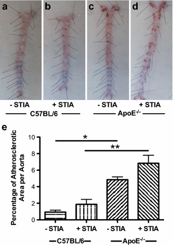
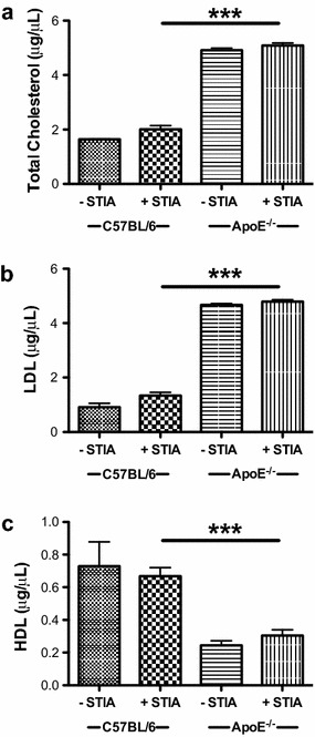
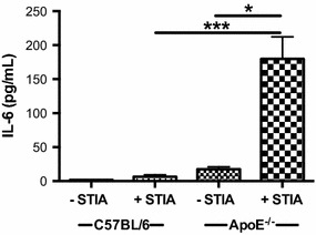
Methods: K/BxN serum transfer-induced arthritis (STIA) was assessed in C57BL/6 (control) and ApoE(-/-) mice using clinical indices and immunohistochemical staining. Ankle synoviums were processed for flow cytometry. Aortic atherosclerosis was quantitated using Sudan IV staining. Serum cholesterol and cytokine levels were determined via enzymatic and luminex bead-based assays, respectively.

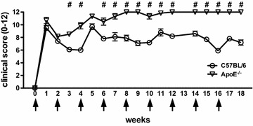
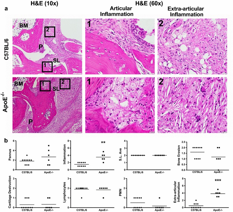
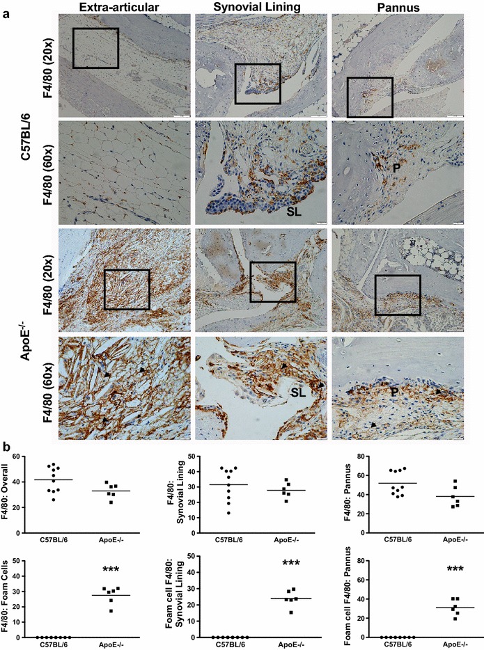
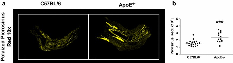
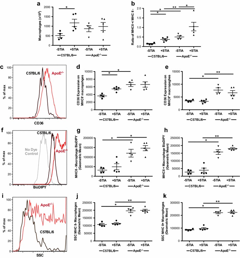
Results: ApoE(-/-) mice developed a sustained and enhanced semi-chronic inflammatory arthritis as compared to control mice. ApoE(-/-) mice had increased numbers of foamy macrophages, enhanced joint inflammation and amplified collagen deposition versus controls. The presence of arthritis did not exacerbate serum cholesterol levels or significantly augment the level of atherosclerosis in ApoE(-/-) mice. However, arthritic ApoE(-/-) mice exhibited a marked elevation of IL-6 as compared to non-arthritic ApoE(-/-) mice and arthritic C57BL/6 mice.

Conclusions: Loss of ApoE potentiates a semi-chronic inflammatory arthritis. This heightened inflammatory response was associated with an increase in circulating IL-6 and in the number of foamy macrophages within the joint. Moreover, the foamy macrophages within the arthritic joint are reminiscent of those within unstable atherosclerotic lesions and suggest a pathologic role for foamy macrophages in propagating arthritis.

Keywords: Animal models of human disease; Arthritis; Cholesterol; Inflammation.


Figures

Fig. 1
Fig. 1
Semi-chronic arthritis is more severe in ApoE−/− mice compared to C57BL/6 mice. C57BL/6 (control) and ApoE−/− mice (n = 4–5 mice/group) were fed chow until 8 weeks of age, at which time they were fed a Western diet for an additional 2–4 months. Animals received serial injections of K/BxN serum every 2–3 weeks, beginning at 8 weeks of age. Arthritis severity was assessed weekly via clinical score. Data are represented as mean ± SEM. Asterisk denotes statistically significant differences: #p < 0.001 for ApoE−/− as compared to control mice. Arrows indicate time of injections
Fig. 2
Fig. 2
Increased articular and extra-articular inflammation is present in ApoE−/− mice. a H&E staining of arthritic ankle joints from C57BL/6 (control, n = 10) and ApoE−/− (n = 6) mice. Boxes 1 and 2 demarcate articular and extra-articular areas of high magnification (×60), respectively. b Histopathological scores for pannus formation, inflammation, synovial lining average, bone erosion, cartilage destruction, lymphocytes, polymorphonuclear cells and extra-articular inflammation on ankle joints from above. Data are represented as mean ± SEM. Asterisk denotes statistically significant differences: **p < 0.01, ***p < 0.001. BM bone marrow, SL synovial lining, P pannus
Fig. 3
Fig. 3
Arthritic ApoE−/− mice have increased F4/80+ foam cells. a Photomicrographs of F4/80-stained extra-articular tissue, synovial lining and pannus of arthritic C57BL/6 (control, n = 10) and ApoE−/− (n = 6) mice. Squares delineate area of higher magnification shown below the respective image. b Immunohistopathological scores for F4/80+ total cells, F4/80+ cells within synovial lining and F4/80+ cells within the pannus from control and ApoE−/−mice. F4/80+ foam cells within each of these categories are subsequently quantified from the mice described above. Data are represented as mean ± SEM. Asterisk denotes statistically significant differences: ***p < 0.001. SL synovial lining, P pannus, arrowheads foamy macrophages
Fig. 4
Fig. 4
Arthritic ApoE−/− mice have increased levels of collagen. a Picrosirius red-positive tissue sections of arthritic C57BL/6 (control, n = 10) and ApoE−/− (n = 6) mice. Picrosirius red staining is pseudocolored yellow. b Quantification of picrosirius red staining on tissue sections (2 sections/mouse) described above. Data are represented as mean ± SEM. Asterisk denotes statistically significant differences: ***p < 0.001
Fig. 5
Fig. 5
Synovial macrophages in ApoE−/− mice have an MHCII+ phenotype and exhibit characteristics of foamy macrophages. Flow cytometric analysis of synovium from 18-week-old non-arthritic and arthritic C57BL/6 (control) and ApoE−/− mice (n = 4–5 mice/group). a Total number of synovial macrophages. b The ratio of MHCII+ to MHC II synovial macrophages. c Histogram of oxidized LDL scavenger receptor, CD36, is shown with concatenation of samples from one experiment of non-arthritic control (black) and ApoE−/− (red) mice. Geometric mean illustrates CD36 expression in d MHCII and e MHCII+ macrophages of individual mice. f Histogram representing uptake of lipophilic dye (BoDIPY) in a concatenation of samples from one experiment of non-arthritic control (black) and ApoE−/− (red) mice. No dye control (grey) is included. Cumulative data displayed as geometric means in g MHCII and h MHCII+ macrophages. i Histogram of side scatter (SSC) of concatenation of samples from one experiment of non-arthritic control (black) and ApoE−/− (red) mice is shown with a composite of samples illustrated as geometric mean of j MHCII and k MHCII+ macrophages. Asterisk denotes statistically significant differences: *p < 0.05, **p < 0.01
Fig. 6
Fig. 6
The presence of arthritis does not affect the development of atherosclerosis in ApoE−/− mice. ad Sudan IV staining of lipids (red) in representative aortic specimens from a, b C57BL/6 (control) and c, d ApoE−/− non-arthritic (a, c) and arthritic mice (b, d). e Quantification of percent area of atherosclerosis (mean % ± SEM) per aorta from control (n = 4 non-arthritic and n = 5 arthritic) and ApoE−/− (n = 4 non-arthritic and n = 5 arthritic) mice at 18 weeks of age. Asterisk denotes statistically significant differences: *p < 0.05, **p < 0.01
Fig. 7
Fig. 7
Serum cholesterol levels are elevated in ApoE−/− mice and remain unchanged during semi-chronic arthritis. Serum levels of a TC, b LDL/vLDL, and c HDL in non-arthritic and arthritic C57BL/6 (control, n = 2 non-arthritic and n = 15 arthritic) and ApoE−/− (n = 3 non-arthritic and n = 6 arthritic) mice at 4–6 months. Data are represented as mean ± SEM. Asterisk denotes statistically significant differences: ***p < 0.001
Fig. 8
Fig. 8
Levels of circulating IL-6 are increased in arthritic ApoE−/− mice. Serum levels of IL-6 from non-arthritic and arthritic C57BL/6 (control, n = 2 non-arthritic and n = 13 arthritic) and ApoE−/− (n = 3 non-arthritic and n = 6 arthritic) mice at 4–6 months. Data are represented as mean ± SEM. Asterisk denotes statistically significant differences. *p < 0.05, ***p < 0.001

References

    1. Bevaart L, Vervoordeldonk MJ, Tak PP. Evaluation of therapeutic targets in animal models of arthritis: how does it relate to rheumatoid arthritis? Arthritis Rheum. 2010;62:2192–2205. doi: 10.1002/art.27503. - DOI - PubMed
    1. Avina-Zubieta JA, Choi HK, Sadatsafavi M, Etminan M, Esdaile JM, Lacaille D. Risk of cardiovascular mortality in patients with rheumatoid arthritis: a meta-analysis of observational studies. Arthritis Rheum. 2008;59:1690–1697. doi: 10.1002/art.24092. - DOI - PubMed
    1. del Rincon I, Williams K, Stern MP, Freeman GL, Escalante A. High incidence of cardiovascular events in a rheumatoid arthritis cohort not explained by traditional cardiac risk factors. Arthritis Rheum. 2001;44:2737–2745. doi: 10.1002/1529-0131(200112)44:12<2737::AID-ART460>3.0.CO;2-#. - DOI - PubMed
    1. Meune C, Touze E, Trinquart L, Allanore Y. Trends in cardiovascular mortality in patients with rheumatoid arthritis over 50 years: a systematic review and meta-analysis of cohort studies. Rheumatology. 2009;48:1309–1313. doi: 10.1093/rheumatology/kep252. - DOI - PubMed
    1. Moore KJ, Sheedy FJ, Fisher EA. Macrophages in atherosclerosis: a dynamic balance. Nat Rev Immunol. 2013;13:709–721. doi: 10.1038/nri3520. - DOI - PMC - PubMed

Publication types

LinkOut - more resources