. 2016 Jul 15;143(14):2522-35.
doi: 10.1242/dev.131797. Epub 2016 Jun 10.

Inhibition of β-catenin signalling in dermal fibroblasts enhances hair follicle regeneration during wound healing

Affiliations

Inhibition of β-catenin signalling in dermal fibroblasts enhances hair follicle regeneration during wound healing

Emanuel Rognoni et al. Development. .

Abstract

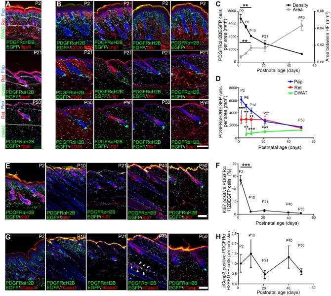
New hair follicles (HFs) do not form in adult mammalian skin unless epidermal Wnt signalling is activated genetically or within large wounds. To understand the postnatal loss of hair forming ability we monitored HF formation at small circular (2 mm) wound sites. At P2, new HFs formed in back skin, but HF formation was markedly decreased by P21. Neonatal tail also formed wound-associated HFs, albeit in smaller numbers. Postnatal loss of HF neogenesis did not correlate with wound closure rate but with a reduction in Lrig1-positive papillary fibroblasts in wounds. Comparative gene expression profiling of back and tail dermis at P1 and dorsal fibroblasts at P2 and P50 showed a correlation between loss of HF formation and decreased expression of genes associated with proliferation and Wnt/β-catenin activity. Between P2 and P50, fibroblast density declined throughout the dermis and clones of fibroblasts became more dispersed. This correlated with a decline in fibroblasts expressing a TOPGFP reporter of Wnt activation. Surprisingly, between P2 and P50 there was no difference in fibroblast proliferation at the wound site but Wnt signalling was highly upregulated in healing dermis of P21 compared with P2 mice. Postnatal β-catenin ablation in fibroblasts promoted HF regeneration in neonatal and adult mouse wounds, whereas β-catenin activation reduced HF regeneration in neonatal wounds. Our data support a model whereby postnatal loss of hair forming ability in wounds reflects elevated dermal Wnt/β-catenin activation in the wound bed, increasing the abundance of fibroblasts that are unable to induce HF formation.

Keywords: Fibroblast lineages; Wounding; β-catenin.


Conflict of interest statement

Competing interests

The authors declare no competing or financial interests.

Figures

Fig. 1.
Fig. 1.
Wound-induced new HF formation in neonatal back and tail skin. (A) Histological analysis of 2 mm wounds performed in PDGFRaH2BeGFP mice postnatally by immunostaining for GFP (green) and Itga6 (red). Arrowheads indicate HFs within the wound bed and white lines demarcate the wound edges. (B) New HF quantitation per wound bed section at PW7. n=4 (P2, P21) or n=3 (other time points) biological replicates. (C) Analysis of different HF morphogenesis stages during HF formation in P2 wounds at PW7. Wound beds were immunostained for GFP (green) and Itga6 (red) or Lef1 (green) alone. (D) Wound-induced new HF formation in tail wounds. Tail wound beds were stained for Krt17 (green) and Itga6 (red) and quantified (see Table S1). Arrowhead indicates new HF. (E-H) Light micrographs and quantification of PW7 wound area in back (E,F) and tail (G,H) skin wounds made at the ages shown. (I) Wound area over time in neonatal and adult wounds as a surrogate for wound closure. n=6 biological replicates. (J-L) Lineage-tracing strategy (J), histological analysis of tdtomato+ fibroblasts (red) labelled with Lrig1CreER and Dlk1CreER (K), and quantitation (L) in PW7 wound beds from P2 wounded mice. (K) Dotted lines denote the epidermal-dermal boundary, arrowheads indicate developing DP and asterisk shows Lrig1CreER-labelled keratinocytes (Page et al., 2013). (L) Percentage of new HFs with attached tdtomato+ fibroblasts. Eight wound bed sections per mouse; n=4 Lrig1CreER, n=3 DLK1CreER. Nuclei are stained with DAPI (blue in A,C,D,K). Data shown are means±s.d. ns, not significant; *P<0.05, **P<0.005, ***P<0.0005. DP, dermal papilla; HF, hair follicle; IP, intraperitoneal. Scale bars: 100 µm in A,D; 50 µm in C,K; 50 mm in E,G.
Fig. 2.
Fig. 2.
Comparative gene expression analysis reveals differences associated with time and body location. (A) Comparison of genes that are differentially expressed between P1 back and tail dermis (location) and between P2 and P50 dorsal fibroblasts (time). Representative GO term analysis of upregulated entities is shown. (B) Heatmap depicting the differentially expressed Wnt target genes among the four samples analysed. Can., canonical; FC, arbitrary fold change; Inv., involved; Neg., negative; Neo., neonatal; Reg., regulation.
Fig. 3.
Fig. 3.
Changes in fibroblast density, marker expression, proliferation and apoptosis in postnatal back skin. (A-D) Fibroblast density and marker expression analysis. Immunostaining for Itga6 (A) and CD26, Lrig1, Dlk1 and Sca1 (red) (B) in PDGFRaH2BeGFP (green) skin. White dotted lines (A) mark dermis layer boundaries. (C) Mean dermal area between adjacent HFs (right y-axis) and total fibroblast density per mm2 of dermis (left y-axis) over time. (D) Fibroblast density in dermis layers indicated in A. (E,F) Ki67 staining (red) of whole-mounts (E) and quantification (F) of percentage Ki67+ PDGFRaH2BeGFP cells (green). (G,H) Cleaved caspase 3 (cCasp3) staining (red) of whole-mounts (G) and quantification (H) of percentage cCasp3+ PDGFRaH2BeGFP cells (green). Note that most apoptotic cells in the skin are observed in the interfollicular epidermis (IFE) and HF keratinocytes (arrowheads in G). Nuclei were labelled with DAPI (blue in A,B,E,G). Pap, papillary dermis; Ret, reticular dermis; DWAT, dermal white adipose tissue. Data shown are means±s.d. n=3 biological replicates per time point (C,D,F,H). **P<0.005, ***P<0.0005. Scale bars: 100 µm.
Fig. 4.
Fig. 4.
Estimation of cellular replication during dermal maturation and clonal analysis of PDGFRaCreERt2-positive cells. (A) Predicted number of dermal fibroblast divisions (trunk skin) during the transition from neonatal (P2) to adult (P50) mouse. Height, length and dermis diameter were measured (n=3 mice per time point and gender) and the dermis volume was estimated by representing the mouse trunk as a cylinder. Cell densities were obtained from Fig. 3C and the cell number at P2 (N0) and P50 (N) were estimated by multiplying cell density and dermis volume. The predicted cell division rate (n=1.3) is calculated by the log2 of the N/N0 ratio. All raw data for the calculations are shown in Table S3. (B-H) Clonal analysis of PDGFRaCreERt2+ cells. (B) Breeding strategy for E-H. (C) Labelling strategy for E,F. (D) Labelling strategy for G,H. (E) Immunostaining of GFP+ fibroblasts (green) co-stained for Dlk1 or Itga8 (red) or overlaid with the brightfield image. (F) Quantitation of GFP+ cells per clone per compartment. n=3 biological replicates per time point. Cells were considered clonally related if they were contained within a total dermal area of 260 µm diameter (Driskell et al., 2013). (G) Immunostaining of GFP+ fibroblasts. (H) Quantitation of GFP+ fibroblasts in the area between adjacent HFs. n=3 biological replicates per time point. Nuclei were labelled with DAPI (blue in E,G). Data shown are means±s.d. Scale bars: 1 cm in A; 100 µm in E (middle and right), G; 500 µm in E (left).
Fig. 5.
Fig. 5.
Dermal Wnt signalling activity. (A,B) Immunostaining for TOPGFP signal (green) and the markers indicated: CD26 (red) and Sca1 (blue) in A; Lef1, TCF1, TCF4, Dkk1 and cyclin D1 (red) in B. Nuclei were labelled with DAPI (white in A, blue in B). (C) Quantitation of percentage TOPGFP+ dermal fibroblasts determined from histological sections. n=3 biological replicates per time point. (D,E) Flow cytometry analysis of TOPGFP expression in P2 fibroblast subpopulations. Gate 1, CD26+ Sca1 (papillary fibroblasts); gate 2, Dlk1+ Sca1 (reticular fibroblasts); gate 3, Dlk1+ Sca1+; gate 4, Dlk1 Sca1+ (cells with adipogenic potential). C is representative of n=3 biological replicates shown in D. Data shown are means±s.d. *P<0.05, ***P<0.0005. Scale bars: 50 µm.
Fig. 6.
Fig. 6.
Analysis of neonatal and adult wound beds. (A,B) TOPGFP expression in fibroblasts of PW5, PW7 and PW10 wound beds of mice wounded at neonatal (P2) and adult (P21 and P50) stages. Wound beds were immunostained for GFP (green), Lrig1 (red) and Sca1 (blue) (A). TOPGFP expression in the boxed areas is shown at higher magnification beneath. Placodes are marked with asterisks and dotted lines indicate the basement membrane. (B) Quantitation of GFP+ cells in wound beds at the indicated time points. n=4 biological replicates, except n=3 for P21 and P50 at PW7. (C) Dermal cells per mm2 wound bed area over time in adult and neonatal wounds. n=4 biological replicates. (D,E) Mean immunofluorescence quantification in the neonatal (P2) and adult (P21, P50) wound beds over time for Lrig1 (D) and Sca1 (E). n=4 biological replicates per time point. (F) Dkk1 expression (red) in TOPGFP (green) P2 and P50 wound beds at PW7. (G,H) PW7 wound beds after wounding at the indicated time point, immunostained for Itga8 (green, G) and αSma (green, H). (I) Quantitation of the density of PDGFRaH2BeGFP-expressing cells in PW7 wound beds wounded at the indicated time points. n=3 biological replicates per time point. (J) Quantitation of Ki67+ cells in the wound beds after wounding at the indicated time points. n=8 (P2), n=6 (P4), n=4 (P21), n=5 (P50) biological replicates. Nuclei were labelled with DAPI (white in A; blue in F-H). Data shown are means±s.d. ns, not significant; *P<0.05, ***P<0.0005. AU, arbitrary units; IF, immunofluorescence. Scale bars: 100 µm.
Fig. 7.
Fig. 7.
Effect of postnatal dermal β-catenin ablation or activation on skin homeostasis. (A,B) Immunostaining of Lrig1 (green, top rows), CD26 (green, bottom rows) and Sca1 (red) in neonatal (P10) and adult (P57) whole-mount sections of wild-type mice (control) or following β-catenin (Ctnnb1) ablation [Ctnnb1(KO)] or activation [Ctnnb1(Ex3)], as shown in Fig. 9A. White dotted lines indicate dermis layer boundaries. (C,D) Quantification of cell densities in the indicated dermis layers in A and B in neonatal (P10, C) and adult (P57, D) whole-mount sections. n=6 whole-mount sections per mouse, n=3 biological replicates per genotype and time point. (E) Quantification of dermal thickness at the indicated time points. n=5 biological replicates per genotype and time point. Nuclei were labelled with DAPI (blue in A,B). Data shown are means±s.d. ns, not significant; *P<0.05, **P<0.005. Scale bars: 100 µm.
Fig. 8.
Fig. 8.
TOPGFP activity and β-catenin deletion efficiency in dermal fibroblasts. (A) TOPGFP activity (green) with Lrig1 immunostaining (red) in P21 wounds at PW7 of wild-type (control) and β-catenin-deleted [Ctnnb1(KO)] mice. (B) Quantitation of GFP+ cells in PW7 wound beds. n=6 whole-mount sections per mouse; n=4 control, n=2 Ctnnb1(KO) biological replicates. (C) Schematic representation of the undeleted and deleted β-catenin targeted allele. DNA from sorted tdtomato-positive (+) and tdtomato-negative (−) cells was tested for β-catenin deletion (primers indicated by red arrows). Ctnnb1(KO) shows a 480 bp band indicating β-catenin deletion and loss of the 180 bp loxP PCR product, whereas undeleted cells (tdtomato) generate only the 180 bp loxP PCR product. In heterozygous mice [Ctnnb1(Het)] the untargeted allele amplifies as a 150 bp band owing to the missing loxP site. Rpl19 PCR was used as loading control. (D) TOPGFP activity (green) with Lrig1 immunostaining (red) in P8 wounds at PW7 of wild-type (control) and β-catenin-stabilised [Ctnnb1(Ex3)] mice. (E) Quantitation of GFP+ cells in PW7 wound beds. n=6 whole-mount sections per mouse; n=4 biological replicates per genotype. (F) Schematic representation of the wild-type, undeleted and deleted targeted exon 3 allele for β-catenin stabilisation. DNA isolated from whole-mount sections was tested for β-catenin exon 3 deletion (primers indicated by red arrows). The wild-type allele amplifies as a 900 bp band, the undeleted targeted allele gives no product and the deleted targeted allele amplifies as a 700 bp band. Note that Ctnnb1(Ex3) transgenic is kept heterozygous, and thus PCR shows efficient recombination. NC, negative control (no DNA). (A,D) Nuclei were labelled with DAPI (blue) and arrowheads indicate new HFs. Data shown are means±s.d. ***P<0.0005. Scale bars: 100 µm.
Fig. 9.
Fig. 9.
Effect of β-catenin ablation or activation on HF formation in wound beds. (A) Experimental design. (B,C) Immunostaining analysis (B) and quantification (C) for Lrig1 (green) and Sca1 (red) in PW7 wound beds of wild-type mice (control) or following β-catenin ablation [Ctnnb1(KO)] or activation [Ctnnb1(Ex3)] wounded at P4. n=3 whole-mount sections per mouse; n=5 biological replicates per genotype. (D-F) Quantification of Ki67+ cells in the wound bed area (D), dermal cells in the wound bed (E) and wound bed area (F) at PW7 of wild-type mice or following β-catenin ablation or activation. Mice were wounded at the indicated time points. (D) n=5 for P50 control, Ctnnb1(KO), n=7 for P4 control, n=4 for P4 Ctnnb1(KO), Ctnnb1 (Ex3), P50 Ctnnb1(Ex3); (E) n=5; (F) n=6 biological replicates. (G,I) Immunostaining of new HFs in PW7 wound beds of mice following β-catenin ablation (G) or activation (I) in postnatal fibroblasts. Wound beds were immunostained for Krt14 (red), Itga6 (green) or tdtomato (red) as indicated. White lines demarcate wound edges; arrowheads indicate new HFs and arrow indicates an enlarged new HF. (H,J) Quantitation of HF formation in wound beds shown in G and I. (H) n=8 for P4 control, P50 Ctnnb1(KO), n=6 for P4 Ctnnb1(KO), n=9 for P50 control; (J) n=9 for P4 control, n=4 for P4 Ctnnb1(Ex3), P50 control, Ctnnb1(Ex3), n=3 for P21 control, Ctnnb1(Ex3) biological replicates. Nuclei were labelled with DAPI (blue in B,G,I). Data shown are means±s.d. ns, not significant; *P<0.05, **P<0.005, ***P<0.0005. AU, arbitrary unit; BW, body weight; IF, immunofluorescence. Scale bars: 100 µm.

References

    1. Andl T., Reddy S. T., Gaddapara T. and Millar S. E. (2002). WNT signals are required for the initiation of hair follicle development. Dev. Cell 2, 643-653. 10.1016/S1534-5807(02)00167-3 - DOI - PubMed
    1. Atit R., Sgaier S. K., Mohamed O. A., Taketo M. M., Dufort D., Joyner A. L., Niswander L. and Conlon R. A. (2006). β-catenin activation is necessary and sufficient to specify the dorsal dermal fate in the mouse. Dev. Biol. 296, 164-176. 10.1016/j.ydbio.2006.04.449 - DOI - PubMed
    1. Betsholtz C. (1995). Role of platelet-derived growth factors in mouse development. Int. J. Dev. Biol. 39, 817-825. - PubMed
    1. Beyer C., Schramm A., Akhmetshina A., Dees C., Kireva T., Gelse K., Sonnylal S., de Crombrugghe B., Taketo M. M., Distler O. et al. (2012). β-catenin is a central mediator of pro-fibrotic Wnt signaling in systemic sclerosis. Ann. Rheum. Dis. 71, 761-767. 10.1136/annrheumdis-2011-200568 - DOI - PMC - PubMed
    1. Breedis C. (1954). Regeneration of hair follicles and sebaceous glands from the epithelium of scars in the rabbit. Cancer Res. 14, 575-579. - PubMed

Publication types

MeSH terms