. 2016 Jun 10;17(1):33.
doi: 10.1186/s12868-016-0270-y.

A mitochondrial division inhibitor, Mdivi-1, inhibits mitochondrial fragmentation and attenuates kainic acid-induced hippocampal cell death

Affiliations

A mitochondrial division inhibitor, Mdivi-1, inhibits mitochondrial fragmentation and attenuates kainic acid-induced hippocampal cell death

Hwajin Kim et al. BMC Neurosci. .

Erratum in

Abstract

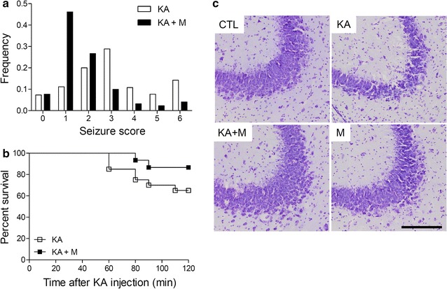
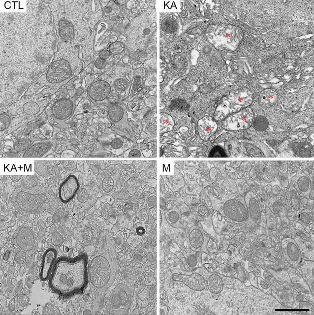
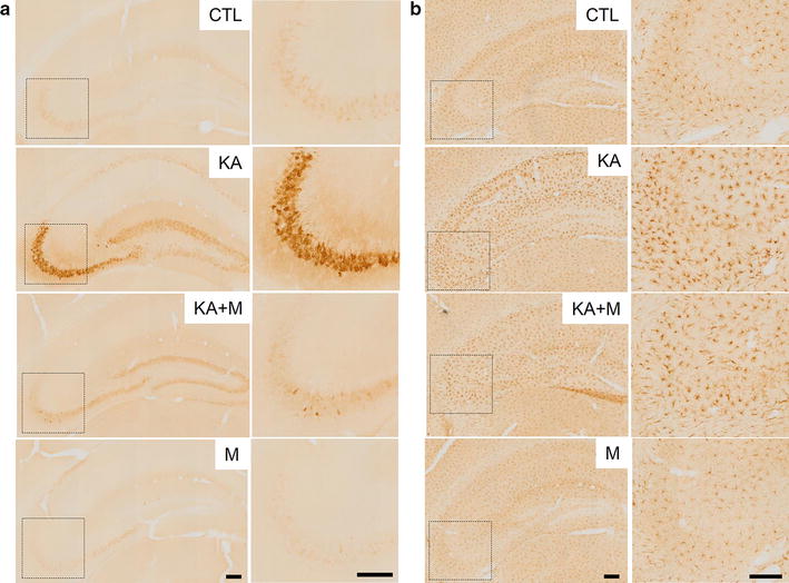
Background: Kainic acid (KA)-induced excitotoxicity promotes cytoplasmic calcium accumulation, oxidative stress, and apoptotic signaling, leading to hippocampal neuronal death. Mitochondria play a critical role in neuroinflammation and the oxidative stress response. Mitochondrial morphology is disrupted during KA-induced seizures; however, it is not clear whether mitochondrial fission or fusion factors are involved in KA-induced neuronal death.

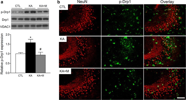
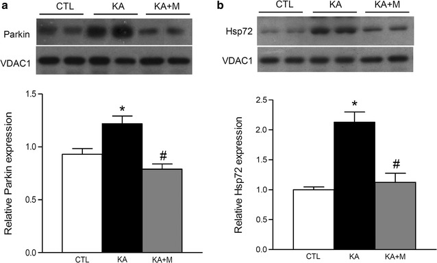
Results: We investigated the effect of Mdivi-1, a chemical inhibitor of the mitochondrial fission protein Drp1, on mitochondrial morphology and function in KA-injected mice. Mdivi-1 pretreatment significantly reduced seizure activity and increased survival rates of KA-treated mice. Mdivi-1 was protective against mitochondrial morphological disruption, and it reduced levels of phosphorylated Drp1 (Ser616) and Parkin recruitment to mitochondria. By contrast, levels of mitochondrial fusion factors did not change. Mdivi-1 also reduced KA-induced neuroinflammation and glial activation.

Conclusions: We conclude that inhibition of mitochondrial fission attenuates Parkin-mediated mitochondrial degradation and protects from KA-induced hippocampal neuronal cell death.

Keywords: Drp1; Mitochondrial fission; Neuroinflammation; Neuronal cell death.


Figures

Fig. 1
Fig. 1
Mdivi-1 effects on KA-induced seizure activity, percent survival, and neuronal cell death. The frequency of behavioral seizure scores (a) and percent survival (b) were monitored during the first 2 h after KA treatment (P < 0.05 KA versus KA + M). c Cresyl violet-stained brain sections showing pyramidal neurons in the CA3 regions. The brain sections were prepared at 24 h after KA injection. Scale bar 200 µm
Fig. 2
Fig. 2
Mdivi-1 effects on mitochondrial morphology in the mouse hippocampus 24 h after KA injection. Representative electron microscopy images showing mitochondrial ultrastructure in the hippocampus of CTL, KA, KA + M, and M mice. Asterisks indicate the mitochondria with disrupted cristae structures. Scale bar 1 µm
Fig. 3
Fig. 3
Mdivi-1 effects on mitochondrial p-Drp1 expression in the mouse hippocampus 24 h after KA injection. a Western blots and quantitative protein analysis of p-Drp1, Drp1, and VDAC1 in the hippocampal mitochondrial fractions from CTL, KA, and KA + M mice. Densitometry values were normalized to Drp1 and expressed as arbitrary units. Data are shown as mean ± SEM. *P < 0.05 versus CTL. # P < 0.05 versus KA. b Representative immunofluorescence images of NeuN, p-Drp1, and overlay in the hippocampus of CTL, KA, and KA + M mice. Scale bar 100 µm
Fig. 4
Fig. 4
Mdivi-1 effects on mitochondrial Parkin and Hsp72 expression in the mouse hippocampus 24 h after KA injection. Western blots and quantitative protein analysis of Parkin (a) and Hsp72 (b) in the hippocampal mitochondrial fractions from CTL, KA, and KA + M mice. Densitometry values were normalized to VDAC1 and expressed as arbitrary units. Data are shown as mean ± SEM. *P < 0.05 versus CTL. # P < 0.05 versus KA
Fig. 5
Fig. 5
Mdivi-1 effects on Cox-2 and Iba-1 expression in the mouse hippocampus 24 h after KA injection. Immunohistochemistry of Cox-2 (a) and Iba-1 (b) in the hippocampus of CTL, KA, KA + M, and M mice. The CA3 regions are indicated as dotted squares and shown magnified at the right. Scale bar 200 µm

References

    1. Korhonen L, Belluardo N, Lindholm D. Regulation of X-chromosome-linked inhibitor of apoptosis protein in kainic acid-induced neuronal death in the rat hippocampus. Mol Cell Neurosci. 2001;17:364–372. doi: 10.1006/mcne.2000.0935. - DOI - PubMed
    1. Olney JW, Rhee V, Ho OL. Kainic acid: a powerful neurotoxic analogue of glutamate. Brain Res. 1974;77:507–512. doi: 10.1016/0006-8993(74)90640-4. - DOI - PubMed
    1. Dong XX, Wang Y, Qin ZH. Molecular mechanisms of excitotoxicity and their relevance to pathogenesis of neurodegenerative diseases. Acta Pharmacol Sin. 2009;30:379–387. doi: 10.1038/aps.2009.24. - DOI - PMC - PubMed
    1. Filosto M, Scarpelli M, Cotelli MS, Vielmi V, Todeschini A, Gregorelli V, Tonin P, Tomelleri G, Padovani A. The role of mitochondria in neurodegenerative diseases. J Neurol. 2011;258:1763–1774. doi: 10.1007/s00415-011-6104-z. - DOI - PubMed
    1. Johri A, Beal MF. Mitochondrial dysfunction in neurodegenerative diseases. J Pharmacol Exp Ther. 2012;342:619–630. doi: 10.1124/jpet.112.192138. - DOI - PMC - PubMed

MeSH terms