. 2016 Dec;6(1):20.
doi: 10.1186/s12348-016-0089-9. Epub 2016 Jun 10.

Bilateral frosted branch angiitis as the presenting sign of antiphospholipid antibody syndrome

Affiliations

Bilateral frosted branch angiitis as the presenting sign of antiphospholipid antibody syndrome

Edward H Wood et al. J Ophthalmic Inflamm Infect. 2016 Dec.

Abstract

Background: "Frosted branch retinal angiitis" is an encompassing term for a rare, typically bilateral diffuse retinal periphlebitis that may occur in a number of varying conditions. To our knowledge, we report the first case of frosted branch angiitis as the presenting sign of antiphospholipid antibody syndrome in a 28-year-old woman.

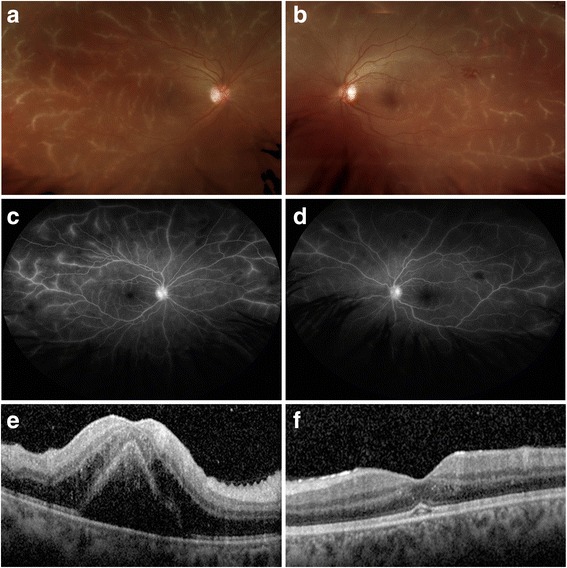
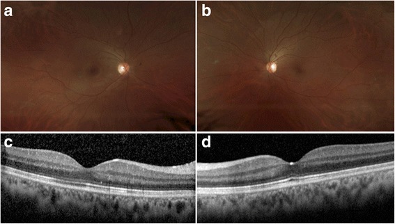
Findings: This study is a retrospective case report and literature review. Serial fundus photos, fluorescein angiogram, and ocular coherence tomography taken were before and after treatment, showing resolution of diffuse retinal perivascular sheathing and macular edema along with marked improvement in visual acuity 4 months after the treatment with corticosteroids.

Conclusions: Frosted branch angiitis can be seen in association with antiphospholipid antibody syndrome. Prompt recognition and treatment with corticosteroids may result in good visual prognosis, and long-term immunosuppression and additional anticoagulation may be beneficial to prevent recurrence.

Keywords: Antiphospholipid antibody syndrome; Frosted branch angiitis; Hydroxychloroquine; Retinal vasculitis.


Figures

Fig. 1
Fig. 1
Fundus photography, fluorescein angiography, and optical coherence tomography of each eye at presentation. a Wide-field fundus photograph of the right eye (OD) showing diffuse retinal periphlebitis. b Wide-field fundus photograph of the left eye (OS) showing diffuse retinal periphlebitis. c Late-phase fluorescein angiogram of the right eye (OD) showing leakage of dye predominantly from the retinal veins and the optic nerve head without deceased transit time or evidence of vascular occlusion. d Late-phase fluorescein angiogram of the left eye (OS) showing leakage of dye predominantly from the retinal veins and the optic nerve head without deceased transit time or evidence of vascular occlusion. e Spectral domain ocular coherence tomography (SD-OCT) through the fovea of the right eye (OD) showing a large amount of cystoid macular edema and a serous foveal detachment. f Spectral domain ocular coherence tomography (SD-OCT) through the fovea of the left eye (OS) showing a smaller foveal detachment with trace intraretinal edema
Fig. 2
Fig. 2
Fundus photography and OCT 4 months after treatment with corticosteroids. a Wide-field fundus photograph of the right eye (OD) showing markedly diminished perivascular sheathing. b Wide-field fundus photograph of the left eye (OS) showing markedly diminished perivascular sheathing. c Spectral domain ocular coherence tomography (SD-OCT) through the fovea of the right eye (OD) showing resolved macular edema. d Spectral domain ocular coherence tomography (SD-OCT) through the fovea of the left eye (OS) showing resolved macular edema

References

    1. Ito Y, Nakano M, Kyu N, Takeuchi M. Frosted branch angiitis in a child. Jpn J Clin Ophthalmol. 1976;30:797–803.
    1. Hernandez-Da Mota SE, Arellanes-Garcia L, Recillas-Gispert C, et al. Lupus relapse presented as frosted branch retinal angiitis: case report. Ocul Immunol Inflamm. 2011;19:367–369. doi: 10.3109/09273948.2011.603879. - DOI - PubMed
    1. Quillen DA, Stathopoulos NA, Blankenship GW, Ferriss JA. Lupus associated frosted branch periphlebitis and exudative maculopathy. Retina. 1997;17:449–451. doi: 10.1097/00006982-199717050-00016. - DOI - PubMed
    1. Kwon SJ, Park DH, Shin JP. Frosted branch angiitis as ocular manifestation of Behcet’s disease: unusual case report and literature review. Korean J Ophthalmol. 2013;27:466–469. doi: 10.3341/kjo.2013.27.6.466. - DOI - PMC - PubMed
    1. Sykes SO, Horton JC. Steroid-responsive retinal vasculitis with a frosted branch appearance in Crohns disease. Retina. 1997;17:451–454. doi: 10.1097/00006982-199717050-00017. - DOI - PubMed

LinkOut - more resources