. 2017 Jan;28(1):85-93.
doi: 10.1681/ASN.2015111282. Epub 2016 Jun 10.

MAP17 Is a Necessary Activator of Renal Na+/Glucose Cotransporter SGLT2

Affiliations

MAP17 Is a Necessary Activator of Renal Na+/Glucose Cotransporter SGLT2

Michael J Coady et al. J Am Soc Nephrol. 2017 Jan.

Abstract

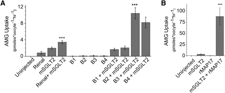
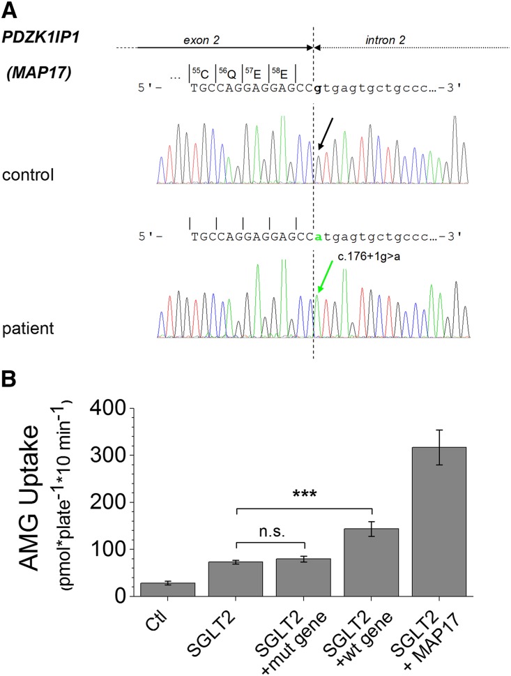
The renal proximal tubule reabsorbs 90% of the filtered glucose load through the Na+-coupled glucose transporter SGLT2, and specific inhibitors of SGLT2 are now available to patients with diabetes to increase urinary glucose excretion. Using expression cloning, we identified an accessory protein, 17 kDa membrane-associated protein (MAP17), that increased SGLT2 activity in RNA-injected Xenopus oocytes by two orders of magnitude. Significant stimulation of SGLT2 activity also occurred in opossum kidney cells cotransfected with SGLT2 and MAP17. Notably, transfection with MAP17 did not change the quantity of SGLT2 protein at the cell surface in either cell type. To confirm the physiologic relevance of the MAP17-SGLT2 interaction, we studied a cohort of 60 individuals with familial renal glucosuria. One patient without any identifiable mutation in the SGLT2 coding gene (SLC5A2) displayed homozygosity for a splicing mutation (c.176+1G>A) in the MAP17 coding gene (PDZK1IP1). In the proximal tubule and in other tissues, MAP17 is known to interact with PDZK1, a scaffolding protein linked to other transporters, including Na+/H+ exchanger 3, and to signaling pathways, such as the A-kinase anchor protein 2/protein kinase A pathway. Thus, these results provide the basis for a more thorough characterization of SGLT2 which would include the possible effects of its inhibition on colocalized renal transporters.

Keywords: Na transport; cell & transport physiology; diabetes; glucose reabsorption; ion transport.


Figures

Figure 1.
Figure 1.
Identification of MAP17 as a factor stimulating SGLT2-mediated AMG uptake. (A) The uptake of AMG into Xenopus oocytes expressing murine SGLT2 mRNA (4.6 ng/oocyte) was stimulated by coexpression of rat renal mRNA (46 ng/oocyte) (the four samples at left; mean±SD; n=5–6 oocytes per sample). When the renal mRNA was size-fractionated, no transport activity was associated with expression of fractions B1–B4 (10 ng/oocyte) but these mRNA samples steeply increased mSGLT2 activity when coinjected with mSGLT2 mRNA, proving that the factor responsible acted by augmenting the activity of the mSGLT2 protein. The Renal+mSGLT2 uptake was significantly different (P<0.001, ANOVA/Bonferroni) from the three other samples shown on the left (Uninjected; Renal; mSGLT2). B3+mSGLT2 uptake is significantly different from mSGLT2 uptake (P<0.001). (B) Expression cloning using a cDNA library produced from mRNA fraction B3 produced a single cDNA clone (rMAP17) whose product very significantly augmented murine SGLT2 activity (P<0.001).
Figure 2.
Figure 2.
Stimulation of human SGLT2 activity with human MAP17 and MARDI. (A) Coexpression of human MAP17 and human SGLT2 in Xenopus oocytes provided results similar to those seen with rat MAP17 and murine SGLT2 (the hSGLT2+hMAP17 uptake was significantly different [P<0.001; mean±SD; n=5–6 oocytes per sample; ANOVA/Bonferroni] from the three other samples, i.e., uninjected, hSGLT2, and hMAP17 mRNA). (B) SGLT2 activity could also be monitored by electrophysiological measurement of the substrate-induced current passing through the protein under voltage-clamp conditions. All traces employ the same scales (as shown). For the first three traces (all from one oocyte held at −50 mV and expressing both hSGLT2 and hMAP17), the changes in current represent the effect of addition of 2 mM glucose (denoted by gray bars) to the bathing solution in the presence of i) no inhibitor; ii) 10 nM dapagliflozin; iii)100 nM dapagliflozin. The other tracings represent the absence of effect of presenting 2 mM glucose to an uninjected oocyte (iv), and oocytes expressing hMAP17 only (v) or hSGLT2 only (vi). (C) Alignment of hMAP17 and MARDI protein sequences. Sequence identity is indicated by white letters on a black background and the two transmembrane (TM) segments are indicated. (D) Coexpression of MARDI causes augmented SGLT2 activity, though less than that seen with MAP17. A typical experiment is shown, of three experiments, where n=6 oocytes for each sample in each experiment. The stimulatory effect of MARDI on SGLT2 expression was significantly different from the effects seen with oocytes expressing either SGLT2 or MARDI alone (P<0.001, ANOVA/Bonferroni).
Figure 3.
Figure 3.
MAP17 activates SGLT2 in transfected OK cells and in oocytes without increasing SGLT2 surface expression. (A) Uptake of radiolabeled AMG into confluent monolayers of OK cells demonstrates that hSGLT2 activity was strongly increased by the presence of hMAP17 (P<0.001) or MAP17Δ where the terminal four amino acids (-STPM) have been removed (P<0.001; mean±SD; n=5 wells per sample; ANOVA/Bonferroni). (B) OK cells expressing both SGLT2 and MAP17 bind more radiolabeled Pz than cells expressing SGLT2 alone (P<0.001; mean±SD; n=5 wells per sample; ANOVA/Bonferroni). Cells expressing SGLT2 alone present a small but significant increase (P<0.05) in Pz binding versus control cells (Ctl). Radiolabeled Pz could be displaced by adding 100 μM cold Pz to the bathing solution. (C) Quantification of superficial immunofluorescent labeling of SGLT2 (flagged with an external F7 epitope) was performed using confluent OK cells fixed with paraformaldehyde. Cells expressing SGLT2 or SGLT2+MAP17 present larger amounts of attached fluorescent antibodies than cells expressing either nothing or MAP17 alone (P<0.01; mean±SD; n=5 wells per sample; ANOVA/Bonferroni). There was no significant difference between SGLT2 and SGLT2+MAP17. (D) OK cells (top panel) were transfected with empty vector (Ctl) or with a vector expressing, F7-hSGLT2 (SGLT2) or F7-hSGLT2 and hMAP17 (SGLT2+MAP17). The amount of primary antibody which had been bound to the cells was observed in a Western blot with a secondary antibody. A similar experiment was performed with plasma membranes isolated from Xenopus oocytes injected with either nothing (Ctl) or mRNAs expressing hSGLT2 or hSGLT2+hMAP17 (n=4). This confirms that the amount of SGLT2 present in the membrane is not increased by the presence of MAP17.
Figure 4.
Figure 4.
The presence of MAP17 does not change the expression level of SGLT2. (A) OK cells expressing pcDNA3.1-F7-hSGLT2 have been fixed with and exposed to an anti-F7 antibody and a fluorescent anti-antibody. (B) OK cells expressing a combination of pcDNA3.1-F7-hSGLT2 and pcDNA3.1-hMAP17-HA. Localization of SGLT2 and similar quantities are visible in both cases. The white scale bars indicate distances of 10 µm and the yellow bar indicates the z axis.
Figure 5.
Figure 5.
Detection of a PDZK1IP1 mutation in a patient with renal glucosuria. (A) Schematic presentation of sequencing results of a PCR product containing exon 2 of the PDZK1IP1gene and an adjacent intronic segment in a patient with FRG. The region around c.176 is shown with the boundary between exon 2 and intron 2 indicated by the dotted line. Note homozygosity for a single base exchange at the first position of the consensus sequence of the donor splice site of intron 2 (c.176+1G>A,IVS2+1G>A) in the patient. (B) Uptake of radiolabeled AMG is shown for OK cells that have been transfected with the vector pcDNA3.1(-) itself (Ctl) or with the recombinant vector containing hSGLT2 cDNA, either alone or cotransfected with the same vector expressing the mutated PDZK1IP1 c.176+1G>A gene (mut gene) or the intact PDZK1IP1 gene (wt gene). For comparison, the AMG uptake obtained from cells cotransfected with the recombinant vectors containing hSGLT2 and hMAP17 is also shown. The AMG uptakes in cells expressing SGLT2 + the mutant MAP17 gene was not different from the uptake in cells expressing SGLT2 alone (P>0.05; mean±SD; n=4 wells; and the entire experiment was repeated three times). A significant difference was observed between the cells cotransfected with the hSGLT2 cDNA and the intact human PDZK1IP1 gene (SGLT2 + wt gene) versus the cells cotransfected with the hSGLT2 cDNA alone (P<0.001, ANOVA/Bonferroni). In addition, the control cells and the cells cotransfected with the two cDNAs were significantly different (P<0.001) from all other samples.

Similar articles

Cited by

References

    1. Sabolic I, Vrhovac I, Eror DB, Gerasimova M, Rose M, Breljak D, Ljubojevic M, Brzica H, Sebastiani A, Thal SC, Sauvant C, Kipp H, Vallon V, Koepsell H: Expression of Na+-D-glucose cotransporter SGLT2 in rodents is kidney-specific and exhibits sex and species differences. Am J Physiol Cell Physiol 302: C1174–C1188, 2012 - PMC - PubMed
    1. Wells RG, Pajor AM, Kanai Y, Turk E, Wright EM, Hediger MA: Cloning of a human kidney cDNA with similarity to the sodium-glucose cotransporter. Am J Physiol 263: F459–F465, 1992 - PubMed
    1. Santer R, Kinner M, Schneppenheim R, Hillebrand G, Kemper M, Ehrich J, Swift P, Skovby F, Schaub J: The molecular basis of renal glucosuria: mutations in the gene for a renal glucose transporter (SGLT2). J Inherit Metab Dis 23[Suppl 1]: 178, 2000
    1. Santer R, Kinner M, Lassen CL, Schneppenheim R, Eggert P, Bald M, Brodehl J, Daschner M, Ehrich JH, Kemper M, Li Volti S, Neuhaus T, Skovby F, Swift PG, Schaub J, Klaerke D: Molecular analysis of the SGLT2 gene in patients with renal glucosuria. J Am Soc Nephrol 14: 2873–2882, 2003 - PubMed
    1. Ullman CG, Frigotto L, Cooley RN: In vitro methods for peptide display and their applications. Brief Funct Genomics 10: 125–134, 2011 - PubMed

Substances

LinkOut - more resources