Review
. 2016 Jun 15;473(12):1681-92.
doi: 10.1042/BCJ20160069.

Control of diverse subcellular processes by a single multi-functional lipid phosphatidylinositol 4,5-bisphosphate [PI(4,5)P2]

Affiliations
Review

Control of diverse subcellular processes by a single multi-functional lipid phosphatidylinositol 4,5-bisphosphate [PI(4,5)P2]

Sourav Kolay et al. Biochem J. .

Abstract

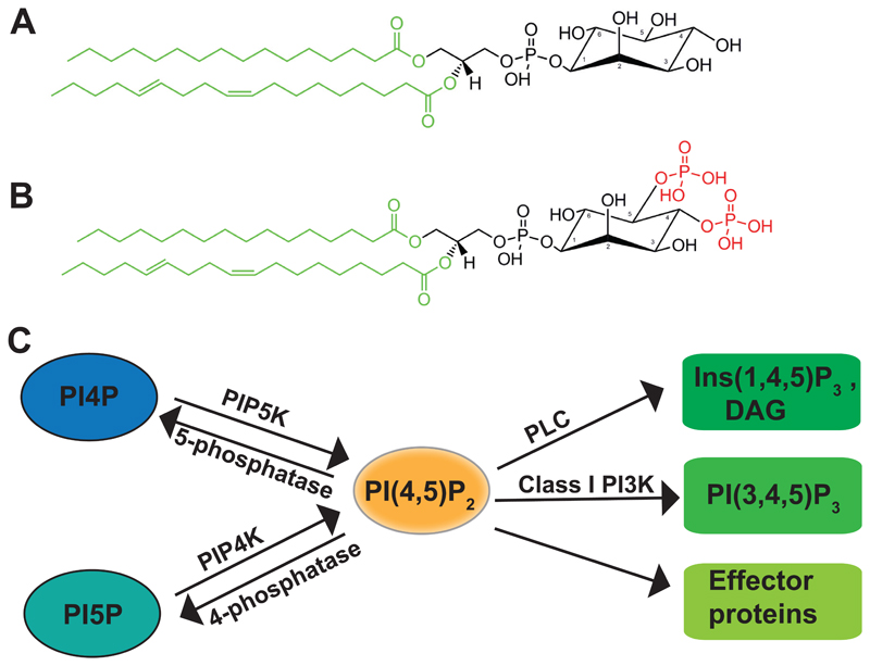
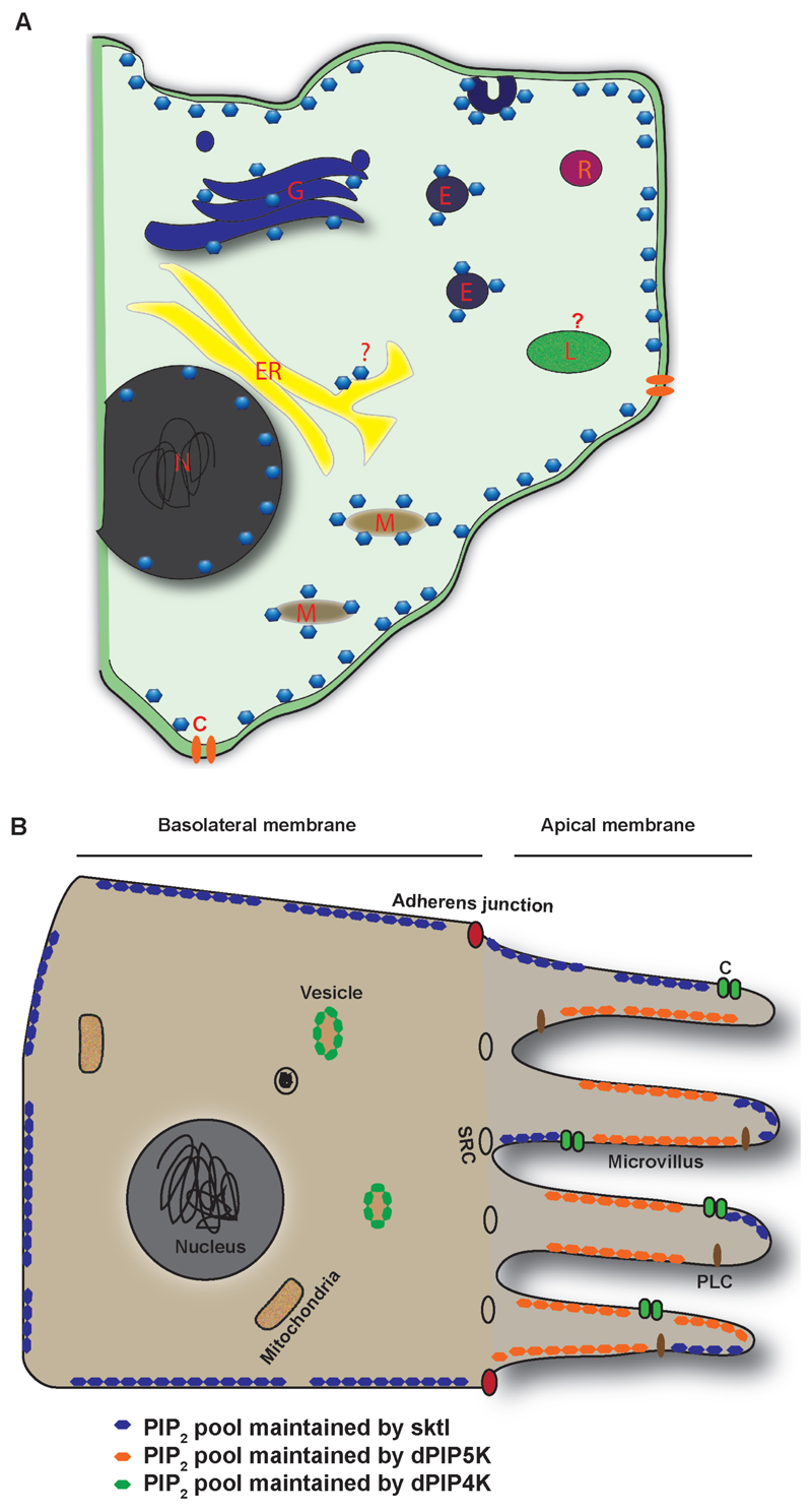
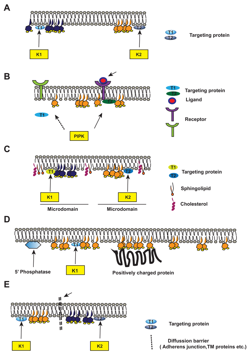
Phosphatidylinositol 4,5-bisphosphate [PI(4,5)P2] is a multi-functional lipid that regulates several essential subcellular processes in eukaryotic cells. In addition to its well-established function as a substrate for receptor-activated signalling at the plasma membrane (PM), it is now recognized that distinct PI(4,5)P2 pools are present at other organelle membranes. However, a long-standing question that remains unresolved is the mechanism by which a single lipid species, with an invariant functional head group, delivers numerous functions without loss of fidelity. In the present review, we summarize studies that have examined the molecular processes that shape the repertoire of PI(4,5)P2 pools in diverse eukaryotes. Collectively, these studies indicate a conserved role for lipid kinase isoforms in generating functionally distinct pools of PI(4,5)P2 in diverse metazoan species. The sophistication underlying the regulation of multiple functions by PI(4,5)P2 is also shaped by mechanisms that regulate its availability to enzymes involved in its metabolism as well as molecular processes that control its diffusion at nanoscales in the PM. Collectively, these mechanisms ensure the specificity of PI(4,5)P2 mediated signalling at eukaryotic membranes.

Keywords: 5)P2 pools; PI(4; cell membranes; lipid kinase; lipid microdomains; phosphoinositides; phospholipase C (PLC) signalling.


Figures

Fig 1
Fig 1. PI(4,5)P2 metabolism pathways
(A) Chemical structure of a molecule of phosphatidylinositol and (B) its phosphorylated derivative phosphatidylinositol 4,5-bisphosphate. The phosphate groups at positions 4 and 5 of the head-group are indicated in red. The acyl chains at sn-1 and sn-2 position that are potential sources of structural and functional diversity in this lipid species are marked in green. (C) Schematic showing the enzymatic pathways of PI(4,5)P2 metabolism; enzymes with known roles in the metabolism of this lipid are shown. PI4P: phosphatidylinositol 4 phosphate, PI5P: phosphatidylinostiol 5-phosphate, PI(4,5)P2: phosphatidylinositol 4,5-bisphosphate, DAG: diacylglycerol, Ins(1,4,5)P3: inositol 1,4,5-trisphosphate, PI(3,4,5)P3: phosphatidylinositol 3,4,5-trisphosphate. PIP5K: phosphatidylinostiol 4-phosphate 5 kinase, PIP4K: phosphatidylinositol 5-phosphate 4 kinase, PLC: phospholipase C, Class 1 PI3K: Class I phosphatidylinositol 3 kinase.
Fig 2
Fig 2. Spatially distributed PI(4,5)2 pools in cells
A) Schematic diagram of a generic cell showing the distribution of PI(4,5)2 pools in various cellular organelles. PI(4,5)P2 is represented as blue hexagonal structures. Cellular organelles and domains are as marked. N: Nucleus, ER: Endoplasmic reticulum, M: Mitochondria, G: Golgi, L: Lysosome, E: Endosome, R: Recycling endosome, C: Ion channel or transporter. B) Schematic of the cross section through a Drosophila photoreceptor cell, depicting the pools of PI(4,5)P2 synthesized by distinct PIPKs in various sub-cellular locations. The apical and basolateral domain of the plasma membrane are indicated. The pools of PI(4,5)P2 are represented in different colors. The cellular organelles and domains are as marked. SRC: sub-rhabdomeric cisternae. PLC depict the distribution of the transduction complex and C the channels at the apical plasma membrane of photoreceptors.
Fig 3
Fig 3. Mechanisms that generate PI(4,5)P2 domains at the plasma membrane
A) Schematic showing two distinct pools of PI(4,5)P2 (hexagonal structures shaded yellow and grey; generated by two separate lipid kinases (K1 and K2); the size of the PI(4,5)P2 headgroup is not scaled). The lipid kinases generate localized pools of PI(4,5)P2. at the membrane. The PM bilayer lipids are shown. Unique proteins (T1, T2) that target K1 and K2 to the PM are shown. B) A model showing receptor activated PI(4,5)P2 synthesis. Activation of a receptor through ligand binding recruits PIP5K through a specific effector proteins T2 that mediates the redistribution of PIP5K to the active signalling receptor and localized PI(4,5)P2 synthesis follows. C) A model depicting two distinct pools of PI(4,5)P2(shaded yellow and grey) generated by K1 and K2 at membrane microdomains such as lipid rafts. The lipid raft region with cholesterol and sphingolipids is shown. The kinases and targeting proteins T1 and T2 are shown. D) Mechanism to restrict the diffusion of a PI(4,5)P2 pool: A model showing localized PI(4,5)P2 production in the PM by the lipid kinase K1 recruited by effector T1. Positively charged proteins at the adjacent plasma membrane sequester the lipid creating a domain of PI(4,5)P2. The localization of a PI(4,5)P2 5-phosphatase activity that limits the boundary of distribution of PI(4,5)P2 generated by K1 is shown. E) A model showing two distinct pools of PI(4,5)P2 (shaded yellow and grey) generated by lipid kinases K1 and K2. The kinases are recruited to plasma membrane by targeting proteins T1 and T2. The dotted line represents a diffusion barrier that prevents free mixing of the PI(4,5)P2 pools generated K1 and K2. The diffusion barrier may be formed by for e.g transmembrane proteins or adherens junction proteins.

References

    1. Pfeffer S, Aivazian D. Targeting Rab GTPases to distinct membrane compartments. Nat Rev Mol Cell Biol. 2004;5:886–96. - PubMed
    1. van Meer G, Voelker DR, Feigenson GW. Membrane lipids: where they are and how they behave. Nat Rev Mol Cell Biol. 2008;9:112–24. - PMC - PubMed
    1. Xu C, Watras J, Loew LM. Kinetic analysis of receptor-activated phosphoinositide turnover. J Cell Biol. 2003;161:779–91. - PMC - PubMed
    1. Jost M, Simpson F, Kavran JM, Lemmon Ma, Schmid SL. Phosphatidylinositol-4,5-bisphosphate is required for endocytic coated vesicle formation. Curr Biol. 8:1399–1402. - PubMed
    1. Ford MG, Pearse BM, Higgins MK, Vallis Y, Owen DJ, Gibson a, Hopkins CR, Evans PR, McMahon HT. Simultaneous binding of PtdIns(4,5)P2 and clathrin by AP180 in the nucleation of clathrin lattices on membranes. Science. 2001;291:1051–1055. - PubMed

Publication types

MeSH terms

Substances