Review
. 2016:76:147-73.
doi: 10.1016/bs.apha.2016.03.002. Epub 2016 Apr 23.

The Long and Winding Road: From the High-Affinity Choline Uptake Site to Clinical Trials for Malignant Brain Tumors

Affiliations
Review

The Long and Winding Road: From the High-Affinity Choline Uptake Site to Clinical Trials for Malignant Brain Tumors

P R Lowenstein et al. Adv Pharmacol. 2016.

Abstract

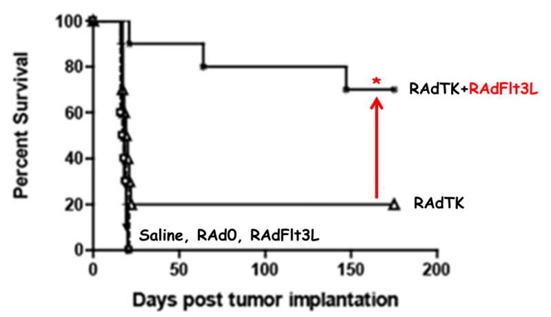
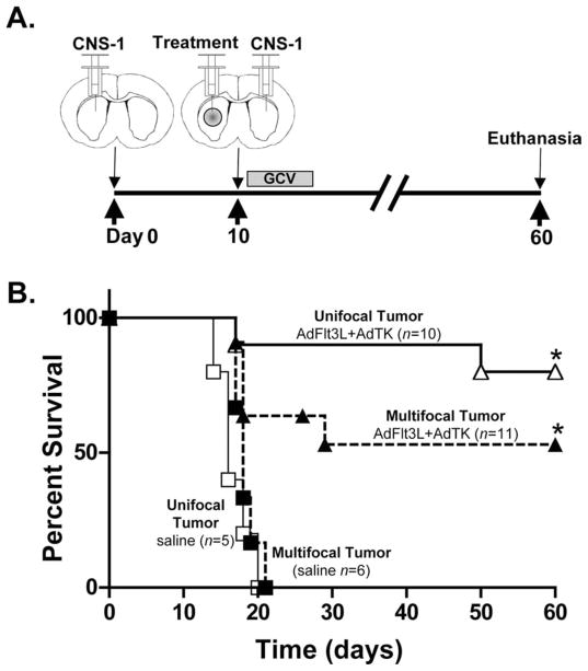
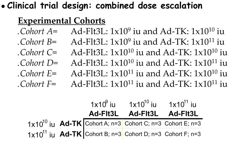
Malignant brain tumors are one of the most lethal cancers. They originate from glial cells which infiltrate throughout the brain. Current standard of care involves surgical resection, radiotherapy, and chemotherapy; median survival is currently ~14-20 months postdiagnosis. Given that the brain immune system is deficient in priming systemic immune responses to glioma antigens, we proposed to reconstitute the brain immune system to achieve immunological priming from within the brain. Two adenoviral vectors are injected into the resection cavity or remaining tumor. One adenoviral vector expresses the HSV-1-derived thymidine kinase which converts ganciclovir into a compound only cytotoxic to dividing glioma cells. The second adenovirus expresses the cytokine fms-like tyrosine kinase 3 ligand (Flt3L). Flt3L differentiates precursors into dendritic cells and acts as a chemokine that attracts dendritic cells to the brain. HSV-1/ganciclovir killing of tumor cells releases tumor antigens that are taken up by dendritic cells within the brain tumor microenvironment. Tumor killing also releases HMGB1, an endogenous TLR2 agonist that activates dendritic cells. HMGB1-activated dendritic cells, loaded with glioma antigens, migrate to cervical lymph nodes to stimulate a systemic CD8+ T cells cytotoxic immune response against glioma. This immune response is specific to glioma tumors, induces immunological memory, and does neither cause brain toxicity nor autoimmune responses. An IND was granted by the FDA on 4/7/2011. A Phase I, first in person trial, to test whether reengineering the brain immune system is potentially therapeutic is ongoing.

Keywords: Brain tumors; Cancer; Flt3L; Gene therapy; Immunotherapy.


Figures

Figure 1
Figure 1
This T1-weighted axial gadolinium-enhanced magnetic resonance image demonstrates an enhancing tumor situated in the left fronto-parietal region. This image is compatible with a high grade malignant glioma (WHO IV). The tumor can be seen as the white amorphous structure to the right of the figure. The darker center represents the tumor proper, with substantial areas of necrosis, while the whitish shadow indicates the enhancement on MRI. Enhancement on MRI suggests impaired blood brain barrier with leakage of contrast agents into the extravascular spaces, i.e. brain edema.
Figure 2
Figure 2
Different brain immune compartments determine systemic immune responses to a viral particulate antigen, influenza hemagglutinin. An adenovirus vector expressing influenza hemagglutinin, RAd-CMV-HA, was injected either into the brain parenchyma (upper left box, blue), into the brain ventricle (upper right box, red), or subcutaneously (lower left box, green). ELISPOT was used to quantify the responses to the influenza hemagglutinin. It can be seen that injections into the brain parenchyma (upper left) did not cause detectable systemic immune responses, while these were seen (as in increased number of spots) when virus was injected into the brain ventricles (upper right). Systemic immune responses were also detected when virus was injected subcutaneously (lower left). As a control, a virus expressing an unrelated protein (β-galactosidase) did not induce an immune response to influenza hemagglutinin.
Figure 3
Figure 3
Schematic illustration of first generation adenoviral vectors (shown above the dotted line), in comparison with the high-capacity helper-dependent adenoviral vectors. Notice that first generation adenoviral vectors retain substantial amounts of viral sequences. Consequently, the packaging limit of the first generation vectors is ~10Kbp, while the high-capacity helper-dependent vectors could carry up to a theoretical value of ~35Kbp. The total size of the adenoviral genome is 36Kbp.
Figure 4
Figure 4
This illustration demonstrates how glioma tumor size determines the response to the therapeutic vector RAdTK + ganciclovir. Syngeneic CNS1 glioma cells were injected into the brains of Lewis rats. A1 shows the size of the tumor at 3 days post-implantation, and B1 shows that if vector therapy is administered at that time 100% of animals are protected from tumor growth. The saline and Radβgal labels indicate that saline injection or a control vector expressing β-galactosidase protein have no therapeutic effect. A3 indicates that if the same treatments are administered to rats at 10 days post tumor implantation, RAdTK + ganciclovir only protect ~15–20% of animals. (Modified from Ali et al., Cancer Research, 65:7194–7204, 2005).
Figure 5
Figure 5
This figure illustrates the effects on tumor progression and animal survival following the addition of RAd-Flt3L. Note that just adding RAd-Flt3L to RAdTK + ganciclovir (as shown in detail in Figure 4) improves long term animal survival from 20% to 77%. The controls for this experiment include saline, RAd0 (an adenovirus vector devoid of transgene), RAd-Flt3L administered alone, and RAd-TK + ganciclovir administered alone. (Modified from Ali et al., Cancer Research, 65:7194–7204, 2005, and, Ali et al., Molecular Therapy, 10:1071–1081–2004)
Figure 6
Figure 6
Treatment of multifocal gliomas with RAdTK + ganciclovir and RAd-Flt3L. A illustrates the experiments schematically. Tumors were implanted first into a single brain hemisphere. Ten days later implanted tumors were treated with gene therapy, and a new tumor was implanted into the contralateral hemisphere, and then followed by ganciclovir. B shows that only treated animals implanted with either a unilateral, or a bilateral tumor survive long term.
Figure 7
Figure 7
Dose-finding Phase I Clinical Trial for the treatment of glioblastoma multiforme. The figure illustrates each of the six experimental cohorts and the amount of each vector which will be injected into the resection cavity of each patient. Below, indicates the combinations of both vectors, and specifically that only two doses of RAd-HSV1-TK will be tested, and three of RAd-Flt3L.

Similar articles

Cited by

References

    1. Abordo-Adesida E, Follenzi A, Barcia C, Sciascia S, Castro MG, Naldini L, Lowenstein PR. Stability of lentiviral vector-mediated transgene expression in the brain in the presence of systemic antivector immune responses. Hum Gene Ther. 2005;16(6):741–751. doi: 10.1089/hum.2005.16.741. - DOI - PMC - PubMed
    1. Ali S, Curtin JF, Zirger JM, Xiong W, King GD, Barcia C, Castro MG. Inflammatory and anti-glioma effects of an adenovirus expressing human soluble Fms-like tyrosine kinase 3 ligand (hsFlt3L): treatment with hsFlt3L inhibits intracranial glioma progression. Mol Ther. 2004;10(6):1071–1084. doi: 10.1016/j.ymthe.2004.08.025. S1525-0016(04)01422-4 [pii] - DOI - PMC - PubMed
    1. Ali S, King GD, Curtin JF, Candolfi M, Xiong W, Liu C, Castro MG. Combined immunostimulation and conditional cytotoxic gene therapy provide long-term survival in a large glioma model. Cancer Res. 2005;65(16):7194–7204. doi: 10.1158/0008-5472.CAN-04-3434. 65/16/7194 [pii] - DOI - PMC - PubMed
    1. Anguille S, Smits EL, Bryant C, Van Acker HH, Goossens H, Lion E, … Berneman ZN. Dendritic Cells as Pharmacological Tools for Cancer Immunotherapy. Pharmacol Rev. 2015;67(4):731–753. doi: 10.1124/pr.114.00945667/4/731. [pii] - DOI - PubMed
    1. Anguille S, Smits EL, Lion E, van Tendeloo VF, Berneman ZN. Clinical use of dendritic cells for cancer therapy. Lancet Oncol. 2014;15(7):e257–267. doi: 10.1016/S1470-2045(13)70585-0. S1470-2045(13)70585-0 [pii] - DOI - PubMed