

Intraoperative cone-beam computed tomography contributes to avoiding hypoglossal nerve palsy during transvenous embolization for dural arteriovenous fistula of the anterior condylar confluence
- PMID: 27288404
- PMCID: PMC5072217
- DOI: 10.1177/1591019916654141
Intraoperative cone-beam computed tomography contributes to avoiding hypoglossal nerve palsy during transvenous embolization for dural arteriovenous fistula of the anterior condylar confluence
Abstract
Background: Dural arteriovenous fistula of the anterior condylar confluence (ACC-DAVF) is a rare subtype of DAVFs that occurs around the hypoglossal canal. Transvenous embolization (TVE) with coils has been performed for most ACC-DAVFs with a high clinical cure rate. However, some reports call attention to hypoglossal nerve palsy associated with TVE due to coil mass compression of the hypoglossal nerve caused by coil deviation from the ACC to the anterior condylar vein (ACV). Herein, we report a case of ACC-DAVF in which an intraoperative cone-beam computed tomography (CT) contributed to avoiding hypoglossal nerve palsy.
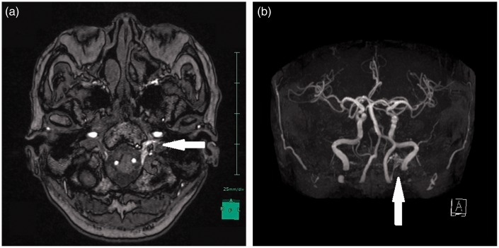
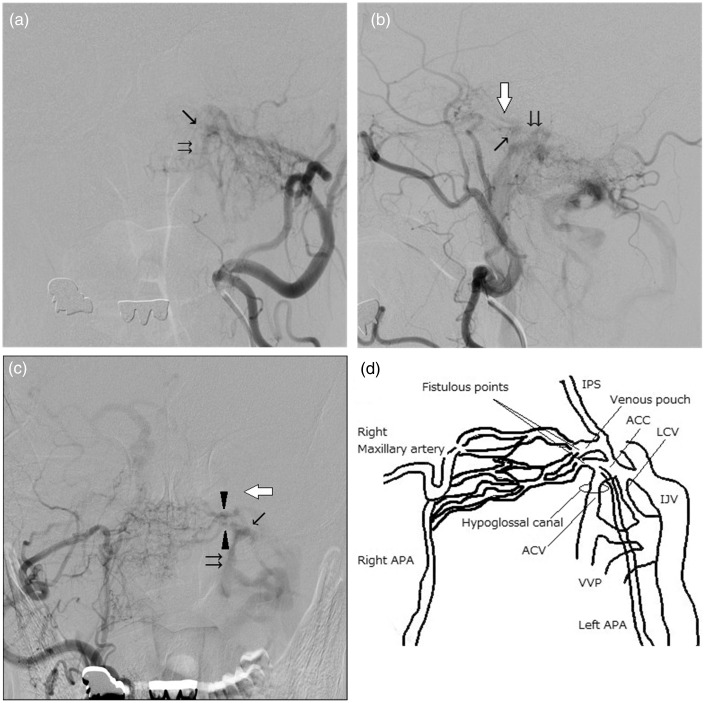
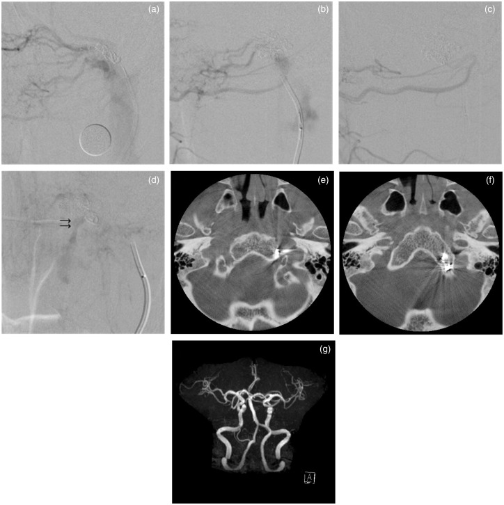
Case presentation: A 74-year-old man presented with left pulse-synchronous tinnitus. An angiography detected left ACC-DAVF mainly supplied by the left ascending pharyngeal artery and mainly drained through the ACV. The two fistulous points were medial side of the ACC and the venous pouch just cranial of the ACC. We performed TVE detecting the fistulous points by contralateral external carotid angiography (ECAG). The diseased venous pouch and ACC were packed with seven coils but a slight remnant of the DAVF was recognized. Because a cone-beam CT revealed that the coil mass was localized in the lateral lower clivus osseous without deviation to the hypoglossal canal, we finished TVE to avoid hypoglossal nerve palsy. Postoperatively, no complication was observed. No recurrence of symptoms or imaging findings were detected during a five-month follow-up period.
Conclusion: An intraoperative cone-beam CT contributed to avoiding hypoglossal nerve palsy by estimating the relationship between the coil mass and the hypoglossal canal during TVE of ACC-DAVF.
Keywords: Anterior condylar confluence; cone-beam computed tomography; dural arteriovenous fistula; hypoglossal nerve palsy; transvenous embolization.
© The Author(s) 2016.
Figures
Similar articles
-
Transvenous coil embolization with intra-operative cone beam CT assistance in the treatment of hypoglossal canal dural arteriovenous fistulae.J Neurointerv Surg. 2019 Feb;11(2):179-183. doi: 10.1136/neurintsurg-2018-014115. Epub 2018 Jul 27. J Neurointerv Surg. 2019. PMID: 30054318
-
Dural arteriovenous fistula of the anterior condylar confluence and hypoglossal canal mimicking a jugular foramen tumor.J Neurosurg. 2008 Aug;109(2):335-40. doi: 10.3171/JNS/2008/109/8/0335. J Neurosurg. 2008. PMID: 18671650
-
Alternative route to a hypoglossal canal dural arteriovenous fistula in case of failed jugular vein approach.Interv Neuroradiol. 2021 Apr;27(2):275-280. doi: 10.1177/1591019920961199. Epub 2020 Oct 7. Interv Neuroradiol. 2021. PMID: 33028133 Free PMC article.
-
Dural arteriovenous fistulas of the hypoglossal canal: systematic review on imaging anatomy, clinical findings, and endovascular management.J Neurosurg. 2015 Apr;122(4):883-903. doi: 10.3171/2014.10.JNS14377. Epub 2014 Nov 21. J Neurosurg. 2015. PMID: 25415064
-
Posterior condylar canal dural arteriovenous fistula: anatomical, symptomatological, and therapeutic considerations in comparison with hypoglossal canal dural arteriovenous fistula.J Neurointerv Surg. 2025 Feb 14;17(3):272-276. doi: 10.1136/jnis-2024-021495. J Neurointerv Surg. 2025. PMID: 38479799 Review.
Cited by
-
Endovascular treatment strategy, technique, and outcomes for dural arteriovenous fistulas of the marginal sinus region.J Neurointerv Surg. 2022 Feb;14(2):155-159. doi: 10.1136/neurintsurg-2021-017476. Epub 2021 May 26. J Neurointerv Surg. 2022. PMID: 34039683 Free PMC article.
-
Morphometric analysis of occipital condyles using alternative imaging technique.Surg Radiol Anat. 2020 Feb;42(2):161-169. doi: 10.1007/s00276-019-02344-2. Epub 2019 Sep 23. Surg Radiol Anat. 2020. PMID: 31549198
-
Dural arteriovenous fistula of the lateral foramen magnum region: A review.Interv Neuroradiol. 2018 Aug;24(4):425-434. doi: 10.1177/1591019918770768. Epub 2018 May 4. Interv Neuroradiol. 2018. PMID: 29726736 Free PMC article. Review.
-
Dural arteriovenous fistulas of the anterior condylar confluence involving the anterior condylar vein within the hypoglossal canal: Two case reports.Surg Neurol Int. 2025 Feb 28;16:69. doi: 10.25259/SNI_7_2025. eCollection 2025. Surg Neurol Int. 2025. PMID: 40041080 Free PMC article.
-
Cone-beam CT-assisted microcatheter tip placement at the shunted pouch entry zone: A technical note for anterior condylar arteriovenous fistula.Neuroradiol J. 2023 Apr;36(2):236-240. doi: 10.1177/19714009221128659. Epub 2022 Sep 20. Neuroradiol J. 2023. PMID: 36124669 Free PMC article.
References
-
- Kuwayama N, Kubo M, Endo S, et al. Present status in the treatment of dural arteriovenous fistulas in Japan. Jpn J Neurosurg (Tokyo) 2011; 20: 12–19.
Publication types
MeSH terms
LinkOut - more resources
Full Text Sources
Other Literature Sources
Miscellaneous
