. 2016 Aug;75(8):731-747.
doi: 10.1093/jnen/nlw045. Epub 2016 Jun 10.

Delayed Hypoxemia Following Traumatic Brain Injury Exacerbates White Matter Injury

Affiliations

Delayed Hypoxemia Following Traumatic Brain Injury Exacerbates White Matter Injury

Umang Parikh et al. J Neuropathol Exp Neurol. 2016 Aug.

Abstract

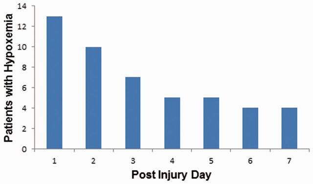
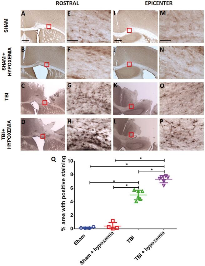
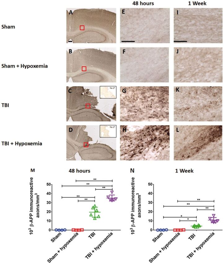
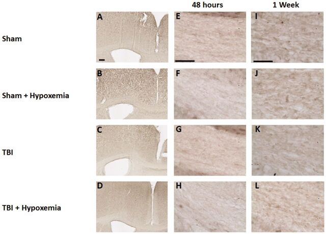
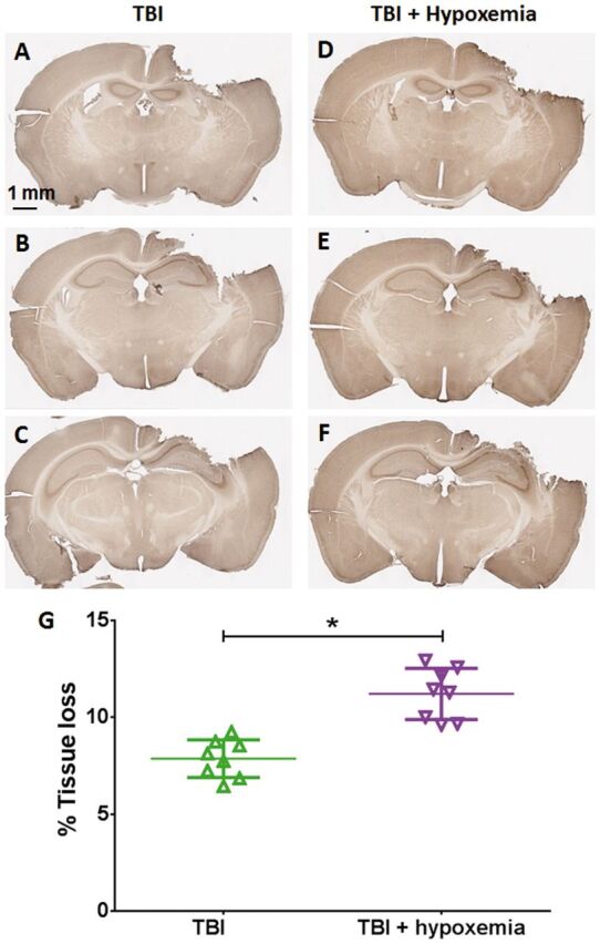
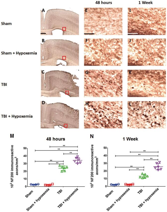
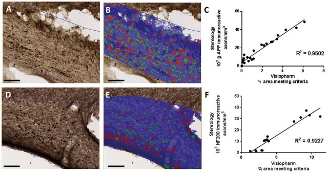
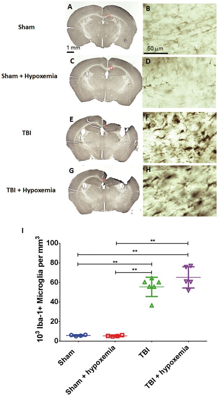
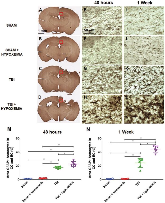
Hypoxemia immediately following traumatic brain injury (TBI) has been observed to exacerbate injury. However, it remains unclear whether delayed hypoxemia beyond the immediate postinjury period influences white matter injury. In a retrospective clinical cohort of children aged 4-16 years admitted with severe TBI, 28/74 (35%) patients were found to experience delayed normocarbic hypoxemia within 7 days of admission. Based on these clinical findings, we developed a clinically relevant mouse model of TBI with delayed hypoxemia by exposing 5-week old (adolescent) mice to hypoxic conditions for 30 minutes starting 24 hours after moderate controlled cortical impact (CCI). Injured mice with hypoxemia had increased axonal injury using both β-amyloid precursor protein and NF200 immunostaining in peri-contusional white matter compared with CCI alone. Furthermore, we detected increased peri-contusional white matter tissue hypoxia with pimonidazole and augmented astrogliosis with anti-glial fibrillary acidic protein staining in CCI + delayed hypoxemia compared with CCI alone or sham surgery + delayed hypoxemia. Microglial activation as evidenced by Iba1 staining was not significantly altered by delayed hypoxemia. These clinical and experimental data indicate the prevention or amelioration of delayed hypoxemia effects following TBI may provide a unique opportunity for the development of therapeutic interventions to reduce axonal injury and improve clinical outcomes.

Keywords: Axonal injury; Brain hypoxia; Controlled cortical impact; Delayed hypoxemia; Secondary injury; Traumatic brain injury.


Figures

FIGURE 1
FIGURE 1
Delayed hypoxemia (defined as O 2 saturation of <90% or PaO 2  <60 mm Hg) in pediatric patients with severe traumatic brain injury (TBI). Frequency and timing of hypoxemia in 74 pediatric patients admitted to the ICU with severe TBI. Forty-eight discreet episodes of hypoxemia were observed in 26 (35%) patients.
FIGURE 2
FIGURE 2
Immunohistochemical staining with the hypoxia marker pimonidazole. (A–D) Pimonidazole staining of white matter rostral to injury epicenter 24 hours after controlled cortical impact (CCI). Scale bar, 250 µm. (E–H) Higher magnification of the white matter. Scale bar, 50 µm. (I–L) Pimonidazole staining white matter at injury epicenter 24 hours after CCI. Scale bar, 250 µm. (M–P) Higher magnification of the white matter. Scale bar, 50 µm. (Q) Image quantification of white matter hypoxia of the ipsilateral corpus callosum and external capsule. *p < 0.0001, ANOVA followed by post hoc Tukey test.
FIGURE 3
FIGURE 3
(A) Hypoxyprobe staining of white matter. (B) Overlay of Visiopharm labeling. Green represents hypoxyprobe staining; blue represents normoxic white matter in the region of interest. Scale bar, 100 µm. (C) Interrater reliability. Visomorph quantification of pimondazole staining of ipsilateral corpus callosum and external capsule by 2 blinded operators. R2  = 0.9953. (D) Test–retest reliability. Visiomorph quantification of pimonidazole staining of ipsilateral corpus callosum and external capsule. Two sets of 12 slices spaced 300 μm apart were stained for pimonidazole binding in the ipsilateral corpus callosum and external capsule and quantified by blinded operators. R2  = 0.9597. Each data point in (C) and (D) represents quantification of 12 slices spaced 300 μm apart from a single brain.
FIGURE 4
FIGURE 4
Controlled cortical impact (CCI) followed by delayed hypoxemia resulted in increased β-APP stained axonal swellings at both 48 hours and 1 week after CCI. (A–D) β-APP staining of the corpus callosum and external capsule at impact epicenter 48 hours postinjury. Scale bar, 250 µm. Insets in (C) and (D) show no primary antibody nonspecific staining at impact epicenter. Scale bar, 250 µm. (E–L) Higher magnification (of red squares in corresponding image) of the white matter at 48 hours (E–H) and 1 week postinjury (I–L) . Scale bar, 50 µm. (M, N) Stereological quantification of β-APP-positive swellings per cubic millimeter of the ipsilateral corpus callosum and external capsule. *p < 0.01, **p < 0.001 ANOVA followed by post hoc Tukey tests.
FIGURE 5
FIGURE 5
β-APP staining of the contralateral corpus callosum or external capsule did not reveal any axonal swellings at either 48 hours or 1 week after controlled cortical impact (CCI). (A–D) β-APP staining of the contralateral white matter at 48 hours postinjury. Scale bar, 250 µm. (E–L) Higher magnification of the contralateral white matter at 48 hours (E–H) and 1 week (I–L) postinjury. Scale bar, 50 µm.
FIGURE 6
FIGURE 6
Tissue loss 1 week after injury. β-APP staining of whole brain slices. (A–F) TBI alone (A–C) and TBI + delayed hypoxemia (D–F) . (G) Quantification of tissue loss of the ipsilateral hemisphere. *p < 0.0001 unpaired t-test.
FIGURE 7
FIGURE 7
Controlled cortical impact (CCI) followed by delayed hypoxemia resulted in increased neurofilament-200 (NF200) stained axonal swellings at both 48 hours and 1 week after CCI. (A–D) NF200 staining of the white matter rostral to impact epicenter at 48 hours postinjury. Scale bar, 250 µm. (E–L) Higher magnification (of red squares in corresponding images) of the white matter at 48 hours (E–H) and 1 week (I–L) postinjury. Scale bar, 50 µm. (M, N) Stereological quantification of NF-200-positive axonal swellings per cubic millimeter of the ipsilateral corpus callosum and external capsule. **p < 0.001, ANOVA followed by post hoc Tukey tests.
FIGURE 8
FIGURE 8
Image quantification of axonal injury strongly correlates with stereological quantification. (A) β-APP staining of ipsilateral corpus callosum and external capsule. (B) Overlay of Visiopharm labeling of β-APP-positive axonal swellings. Green represents axonal swellings in the short axis; red represents axonal swellings in the long axis. Scale bar, 100 µm. (C) Correlation of stereological quantification and Visiopharm image analysis of ipsilateral corpus callosum and external capsule. R2  = 0.9502. Each data point represents quantification of 12 slices spaced 300 μm apart from 1 brain 48 hours or 1 week after injury or sham surgery. (D) NF200 staining of ipsilateral corpus callosum and external capsule. (E) Overlay of Visiopharm labeling of β-APP-positive axonal swellings. Green represents axonal swellings in the short axis; red represents axonal swellings in the long axis. Scale bar, 100 µm. (F) Correlation of stereological quantification and Visiopharm image analysis of ipsilateral corpus callosum and external capsule. R2  = 0.9227. Each data point represents quantification of 12 slices spaced 300 μm apart from 1 brain 48 hours or 1 week after injury or sham surgery.
FIGURE 9
FIGURE 9
Microglial activation 1 week after traumatic brain injury (TBI). (A–H) Iba-1 stained whole sections (A, C, E, G) and higher magnification of the ipsilateral corpus callosum (B, D, F, H) at 1 week postinjury. (I) Stereological quantification of Iba-1-positive microglia in the ipsilateral corpus callosum and external capsule did not demonstrate a statistically significant difference between the 2 injury groups. **p < 0.001, ANOVA followed by post hoc Tukey tests.
FIGURE 10
FIGURE 10
Increased astrocytosis following delayed hypoxemia after traumatic brain injury (TBI). (A–L) Anti-GFAP stained whole sections 48 hours after TBI (A–D) . Higher magnification of the corpus callosum (red squares in corresponding images) 48 hours after TBI (E–H) and 1 week after TBI (I–L) . (M, N) Percent area of GFAP staining in the ipsilateral corpus callosum (CC) and external capsule (EC). *p < 0.01, **p < 0.001 ANOVA followed by post hoc Tukey tests.

Similar articles

Cited by

References

    1. Heron M, Sutton PD, Xu J , et al. . Annual summary of vital statistics: 2007 . Pediatrics 2010. ; 125 : 4 – 15 - PubMed
    1. Fisher MD. Pediatric traumatic brain injury . Crit Care Nurs 1997. ; 20 : 36 – 51 - PubMed
    1. Kochanek PM, Clark RS, Ruppel RA , et al. . Biochemical, cellular, and molecular mechanisms in the evolution of secondary damage after severe traumatic brain injury in infants and children: Lessons learned from the bedside . Pediatr Crit Care Med 2000. ; 1 : 4 – 19 - PubMed
    1. Chesnut RM, Marshall LF, Klauber MR , et al. . The role of secondary brain injury in determining outcome from severe head injury . J Trauma 1993. ; 34 : 216 – 22 - PubMed
    1. Chi JH, Knudson MM, Vassar MJ , et al. . Prehospital hypoxia affects outcome in patients with traumatic brain injury: A prospective multicenter study . J Trauma 2006. ; 61 : 1134 – 41 - PubMed