. 2016 Jun 13:7:ncomms11836.
doi: 10.1038/ncomms11836.

Cross-membranes orchestrate compartmentalization and morphogenesis in Streptomyces

Affiliations

Cross-membranes orchestrate compartmentalization and morphogenesis in Streptomyces

Katherine Celler et al. Nat Commun. .

Abstract

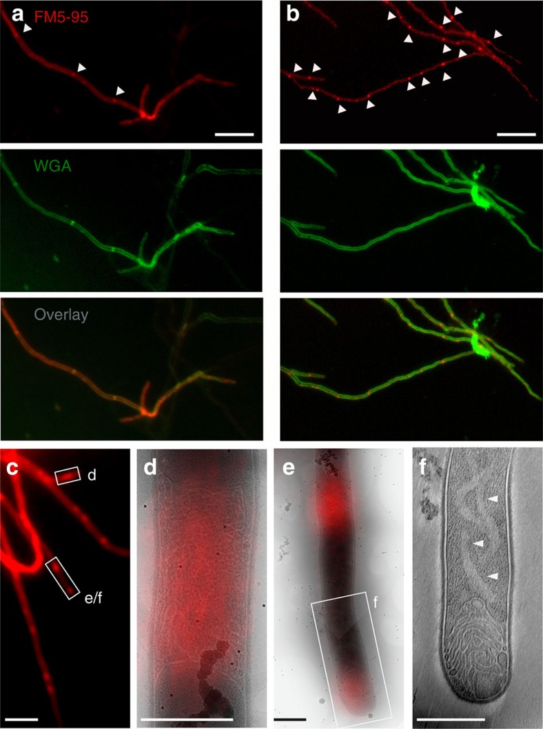
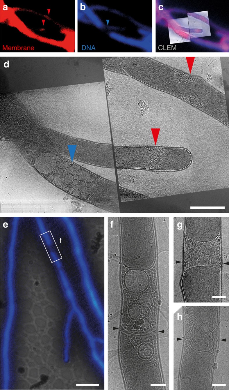
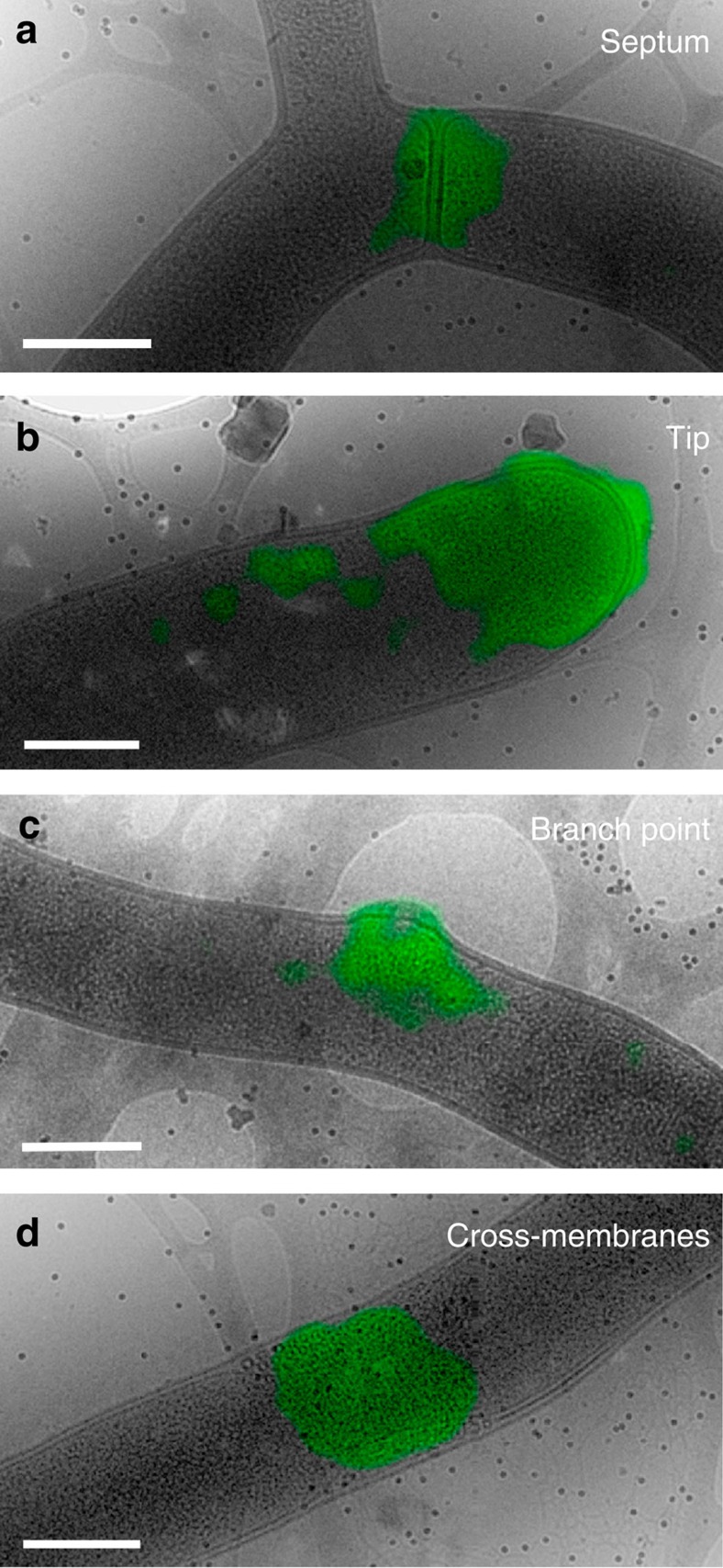
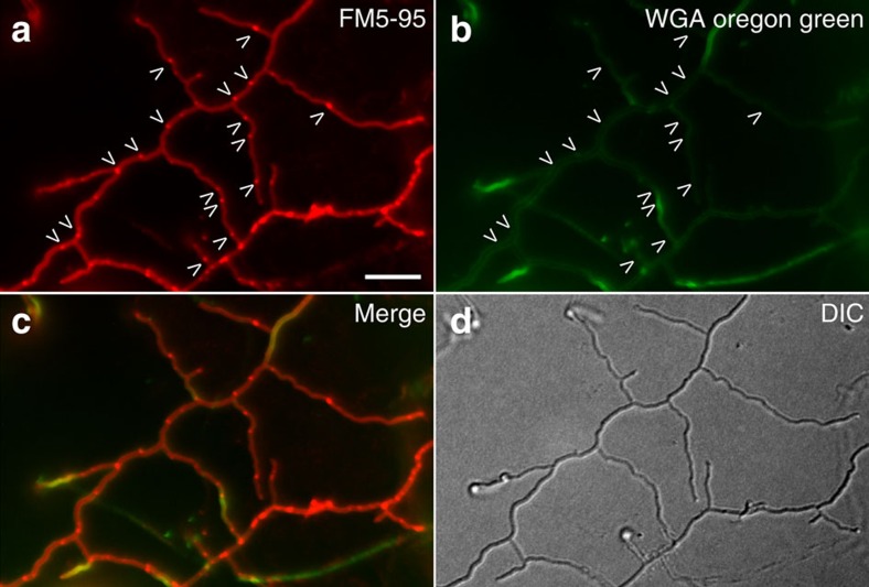
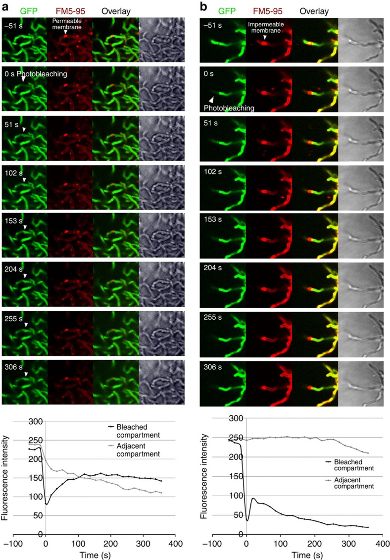
Far from being simple unicellular entities, bacteria have complex social behaviour and organization, living in large populations, and some even as coherent, multicellular entities. The filamentous streptomycetes epitomize such multicellularity, growing as a syncytial mycelium with physiologically distinct hyphal compartments separated by infrequent cross-walls. The viability of mutants devoid of cell division, which can be propagated from fragments, suggests the presence of a different form of compartmentalization in the mycelium. Here we show that complex membranes, visualized by cryo-correlative light microscopy and electron tomography, fulfil this role. Membranes form small assemblies between the cell wall and cytoplasmic membrane, or, as evidenced by FRAP experiments, large protein-impermeable cross-membrane structures, which compartmentalize the multinucleoid mycelium. All areas containing cross-membrane structures are nucleoid-restricted zones, suggesting that the membrane assemblies may also act to protect nucleoids from cell-wall restructuring events. Our work reveals a novel mechanism of controlling compartmentalization and development in multicellular bacteria.


Figures

Figure 1
Figure 1. Membrane assemblies in vegetative hyphae.
(a) In vegetative hyphae of S. coelicolor M145 stained with the membrane dye FM5–95 (red) and the cell wall dye FITC-WGA (green), cross-walls are evident (arrowheads). (b) Large membrane structures/agglomerates are also found within hyphae, which do not co-localize with WGA-stained cross-walls (arrowheads). (cf) To investigate their ultrastructure, membranes in S. albus were fluorescently labelled with FM5–95 and imaged with cryo-CLEM. Positions with extended lipids were observed (df). Cross-membrane assemblies within hyphae (d,e) and at tips (e,f) consisted of extended tubular membrane structures. A light region, devoid of ribosomes and other macromolecular complexes, can be seen forming a faint ribbon behind a membrane-filled tip (arrows, f; for tomogram see Supplementary Movie 1). Scale bars, 5 μm (ac), 500 nm (df).
Figure 2
Figure 2. Electron tomography and data segmentation of membrane formations in three dimensions.
Cryo-ET slices (top row), surface renderings of the tomograms (middle row) and 2D model (bottom rows, side view and top view) of intracellular membranes in vegetative hyphae of Streptomyces. When no membranes are observed, hyphae appear as in a. An example of small patches, or blebs, of densely packed lipid tubes between the cytoplasmic membrane and cell wall is given in b. White arrowheads denote lighter areas, devoid of ribosomes. In c, lipid tubes can be seen increasingly constricting the cytoplasm, whereas in d, full cross-membranes are evident, forming a plug across the hypha. In the 2D model, cell wall (red), DNA (dark blue), cytoplasm (light blue), cytoplasmic membrane (black), membrane tubes and vesicles (green) are schematically depicted from orthogonal (third row) and parallel views (fourth row).
Figure 3
Figure 3. DNA and cross-membranes are mutually exclusive in vegetative hyphae.
Vegetative hyphae of S. albus fluorescently labelled with FM5–95 for membranes (a) and DAPI for DNA (b) were imaged by cryo-CLEM (c) and cryo-ET (d). All areas that contained membrane assemblies (red arrowheads, a,d) were devoid of DNA. Vesicularization could be seen near a completed cross-wall in an area devoid of DNA (blue arrowheads, b,d). In a second example, fluorescent labelling of vegetative hyphae of S. albus with DAPI (e) again revealed positions devoid of DNA (white rectangle denoted f) and filled with membranes and vesicles as shown by cryo-EM. Cell wall formation can be seen within the membranes (black arrowheads, f). In g,h, other examples of newly forming cell wall within membranes are shown. Scale bars, 500 nm ad; e, 5 μm; fh, 200 nm.
Figure 4
Figure 4. Vancomycin-BODIPY staining shows positions of cell wall restructuring.
Cryo-CLEM on vegetative hyphae of S. albus that were fluorescently labelled with vancomycin-BODIPY showed that restructuring of cell wall mainly occurs at sites of septa (a), but also at tips (b), branch points (c) and cross-membranes (d).
Figure 5
Figure 5. Membrane and cell wall localization in an S. coelicolor ftsZ-deletion strain.
Membrane assemblies are still abundant in S. coelicolor ftsZ-deletion strain (arrows, a, stained with FM5–95), but no cross-walls form (arrows, b, note N-acetylglucosaminyl residues present in cell wall stained with WGA-Oregon green). An overlay image is provided (c), as well as the light image (d). Scale bar, 5 μm. DIC, differential interference contrast.
Figure 6
Figure 6. FRAP analysis of GFP distribution over cross-membranes.
Fluorescence recovery after photobleaching (FRAP) on a S. coelicolor strain constitutively expressing eGFP and stained with FM5–95 for membranes. An entire compartment was bleached on one side of a cross-membrane to establish whether GFP molecules from the adjacent compartment would restore fluorescence. Membranes can be both cytosol permeable (a and Supplementary Movie 6), which can be seen when bleached GFP molecules spread through the membrane as indicated by the white arrows, or impermeable (b and Supplementary Movie 7), which can be seen when the membrane prevents the flow of bleached GFP molecules. Recovery curves demonstrate the recovery in fluorescence intensity in the case of the permeable membrane, together with a decrease in fluorescence in the adjacent compartment. In the case of the impermeable membrane, a slight increase in fluorescence is seen in the bleached compartment due to diffusion of GFP molecules from within the compartment, whereas there is no decrease in fluorescence in the adjacent compartment. Further imaging results in bleaching and a decrease in fluorescence in both the bleached and adjacent compartments.

References

    1. Shapiro J. A. Thinking about bacterial populations as multicellular organisms. Annu. Rev. Microbiol. 52, 81–104 (1998). - PubMed
    1. Claessen D., Rozen D. E., Kuipers O. P., Sogaard-Andersen L. & van Wezel G. P. Bacterial solutions to multicellularity: a tale of biofilms, filaments and fruiting bodies. Nat. Rev. Microbiol. 12, 115–124 (2014). - PubMed
    1. Lyons N. A. & Kolter R. On the evolution of bacterial multicellularity. Curr. Opin. Microbiol. 24, 21–28 (2015). - PMC - PubMed
    1. Miguelez E. M., Hardisson C. & Manzanal M. B. Hyphal death during colony development in Streptomyces antibioticus: morphological evidence for the existence of a process of cell deletion in a multicellular prokaryote. J. Cell Biol. 145, 515–525 (1999). - PMC - PubMed
    1. Miguelez E. M., Hardisson C. & Manzanal M. B. Streptomycetes: a new model to study cell death. Int. Microbiol. 3, 153–158 (2000). - PubMed

Publication types

MeSH terms