

Evolutionary and developmental analysis reveals KANK genes were co-opted for vertebrate vascular development
- PMID: 27292017
- PMCID: PMC4904190
- DOI: 10.1038/srep27816
Evolutionary and developmental analysis reveals KANK genes were co-opted for vertebrate vascular development
Abstract
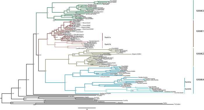
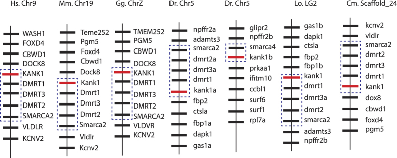
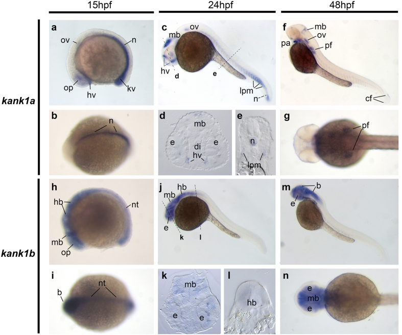
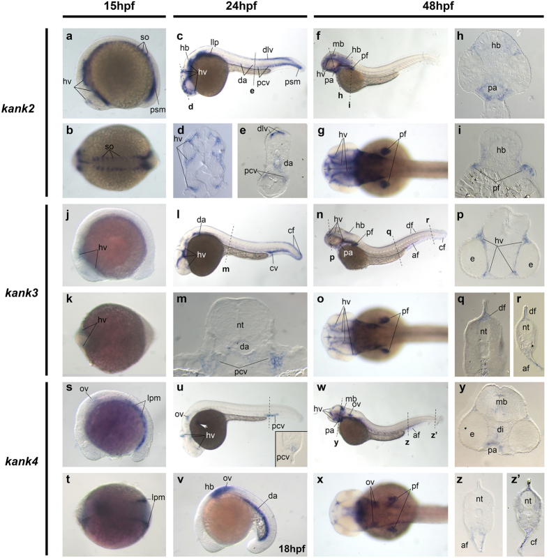
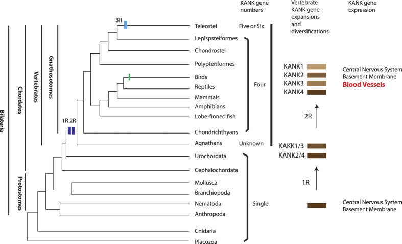
Gene co-option, usually after gene duplication, in the evolution of development is found to contribute to vertebrate morphological innovations, including the endothelium-based vascular system. Recently, a zebrafish kank gene was found expressed in the vascular vessel primordium, suggesting KANK genes are a component of the developmental tool kit for the vertebrate vascular system. However, how the KANK gene family is involved in vascular vessel development during evolution remains largely unknown. First, we analyzed the molecular evolution of the KANK genes in metazoan, and found that KANK1, KANK2, KANK3 and KANK4 emerged in the lineage of vertebrate, consistent with the two rounds of vertebrate whole-genome duplications (WGD). Moreover, KANK genes were further duplicated in teleosts through the bony-fish specific WGD, while only kank1 and kank4 duplicates were retained in some of the examined fish species. We also found all zebrafish kank genes, except kank1b, are primarily expressed during embryonic vascular development. Compared to invertebrate KANK gene expression in the central nervous system, the vascular expression of zebrafish kank genes suggested KANK genes were co-opted for vertebrate vascular development. Given the cellular roles of KANK genes, our results suggest that this co-option may facilitate the evolutionary origin of vertebrate vascular vessels.
Figures
Similar articles
-
Expression patterns of dscam and sdk gene paralogs in developing zebrafish retina.Mol Vis. 2018 Jul 19;24:443-458. eCollection 2018. Mol Vis. 2018. PMID: 30078982 Free PMC article.
-
Kank proteins: a new family of ankyrin-repeat domain-containing proteins.Biochim Biophys Acta. 2008 Feb;1780(2):128-33. doi: 10.1016/j.bbagen.2007.09.017. Epub 2007 Oct 4. Biochim Biophys Acta. 2008. PMID: 17996375
-
The evolutionary conservation of the A Disintegrin-like and Metalloproteinase domain with Thrombospondin-1 motif metzincins across vertebrate species and their expression in teleost zebrafish.BMC Evol Biol. 2015 Feb 15;15:22. doi: 10.1186/s12862-015-0281-9. BMC Evol Biol. 2015. PMID: 25879701 Free PMC article.
-
The zebrafish genome in context: ohnologs gone missing.J Exp Zool B Mol Dev Evol. 2007 Sep 15;308(5):563-77. doi: 10.1002/jez.b.21137. J Exp Zool B Mol Dev Evol. 2007. PMID: 17068775 Review.
-
Impact of gene gains, losses and duplication modes on the origin and diversification of vertebrates.Semin Cell Dev Biol. 2013 Feb;24(2):83-94. doi: 10.1016/j.semcdb.2012.12.008. Epub 2013 Jan 3. Semin Cell Dev Biol. 2013. PMID: 23291262 Review.
Cited by
-
Proteomic profiling of concurrently isolated primary microvascular endothelial cells, pericytes, and vascular smooth muscle cells from adult mouse heart.Sci Rep. 2022 May 25;12(1):8835. doi: 10.1038/s41598-022-12749-6. Sci Rep. 2022. PMID: 35614104 Free PMC article.
-
Evolutionary and Expression Analyses Show Co-option of khdrbs Genes for Origin of Vertebrate Brain.Front Genet. 2018 Jan 4;8:225. doi: 10.3389/fgene.2017.00225. eCollection 2017. Front Genet. 2018. PMID: 29354154 Free PMC article.
-
Extreme-Phenotype Genome-Wide Association Analysis for Growth Traits in Spotted Sea Bass (Lateolabrax maculatus) Using Whole-Genome Resequencing.Animals (Basel). 2024 Oct 17;14(20):2995. doi: 10.3390/ani14202995. Animals (Basel). 2024. PMID: 39457925 Free PMC article.
-
Phylogenetic and developmental analyses indicate complex functions of calcium-activated potassium channels in zebrafish embryonic development.Dev Dyn. 2021 Oct;250(10):1477-1493. doi: 10.1002/dvdy.329. Epub 2021 Mar 24. Dev Dyn. 2021. PMID: 33728688 Free PMC article.
-
A germline chimeric KANK1-DMRT1 transcript derived from a complex structural variant is associated with a congenital heart defect segregating across five generations.Res Sq [Preprint]. 2023 Dec 13:rs.3.rs-3740005. doi: 10.21203/rs.3.rs-3740005/v1. Res Sq. 2023. Update in: Chromosome Res. 2024 Mar 19;32(2):6. doi: 10.1007/s10577-024-09750-2. PMID: 38168413 Free PMC article. Updated. Preprint.
References
-
- Holland P. W., Garcia-Fernandez J., Williams N. A. & Sidow A. Gene duplications and the origins of vertebrate development. Dev Suppl, 125–133 (1994). - PubMed
-
- Carroll S. B. Endless forms: the evolution of gene regulation and morphological diversity. Cell 101, 577–580 (2000). - PubMed
-
- Ohno S. Evolution by Gene Duplication. (Springer Science+Business Media, LLC, 1970).
Publication types
MeSH terms
Substances
Grants and funding
LinkOut - more resources
Full Text Sources
Other Literature Sources
Molecular Biology Databases
Research Materials
