. 2016 Jun 13:7:ncomms11852.
doi: 10.1038/ncomms11852.

Long-lived self-renewing bone marrow-derived macrophages displace embryo-derived cells to inhabit adult serous cavities

Affiliations

Long-lived self-renewing bone marrow-derived macrophages displace embryo-derived cells to inhabit adult serous cavities

Calum C Bain et al. Nat Commun. .

Abstract

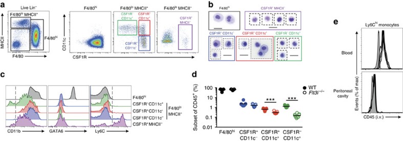
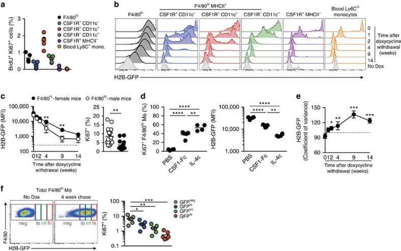
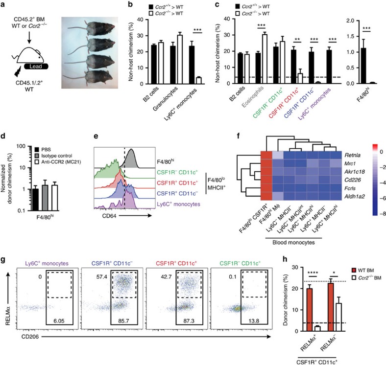
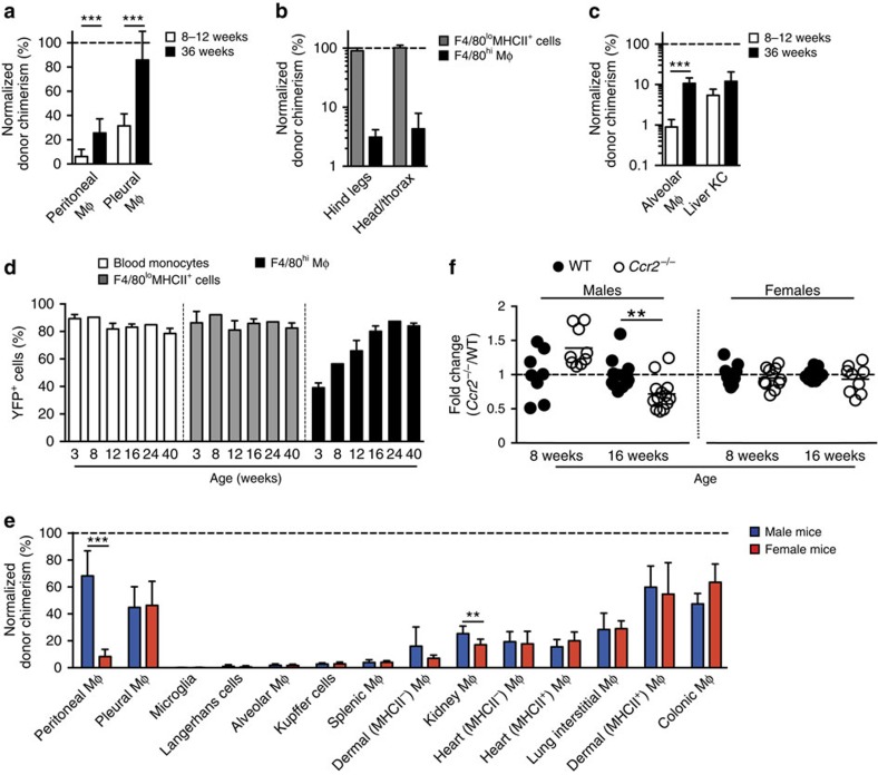
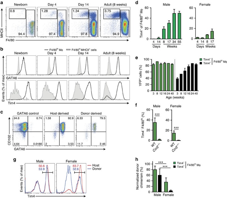
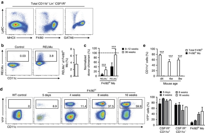
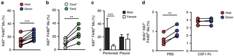
Peritoneal macrophages are one of the most studied macrophage populations in the body, yet the composition, developmental origin and mechanisms governing the maintenance of this compartment are controversial. Here we show resident F4/80(hi)GATA6(+) macrophages are long-lived, undergo non-stochastic self-renewal and retain cells of embryonic origin for at least 4 months in mice. However, Ly6C(+) monocytes constitutively enter the peritoneal cavity in a CCR2-dependent manner, where they mature into short-lived F4/80(lo)MHCII(+) cells that act, in part, as precursors of F4/80(hi)GATA6(+) macrophages. Notably, monocyte-derived F4/80(hi) macrophages eventually displace the embryonic population with age in a process that is highly gender dependent and not due to proliferative exhaustion of the incumbent embryonic population, despite the greater proliferative activity of newly recruited cells. Furthermore, although monocyte-derived cells acquire key characteristics of the embryonic population, expression of Tim4 was impaired, leading to cumulative changes in the population with age.


Figures

Figure 1
Figure 1. Phenotypic characterization of peritoneal myeloid cells.
(a) Expression of F4/80 and MHCII by live CD45+Lin (CD3, CD19, Ly6G, SiglecF) peritoneal exudate cells (PEC) obtained from naïve adult WT mice (left panel) and expression of CD11c and CSF1R by F4/80hi, F4/80loMHCII+ and F4/80loMHCII cells (right panel). Data are representative of at least 20 independent experiments. (b) Morphological appearance of F4/80hi, F4/80loMHCII+ subsets and F4/80loMHCII cells purified from naive adult WT mice. Scale bar, 20 μm. Images are from one experiment performed. (c) Expression of CD11b, GATA6 and Ly6C by PEC myeloid subsets as gated in a. Histograms are representative of at least six (GATA6) or 20 (CD11b and Ly6C) independent experiments. (d) Frequency among CD45+ leukocytes of F4/80hi macrophages and CD11c/CSF1R-defined F4/80loMHCII+ subsets in PEC of naive 8-week old WT (n=9) or Flt3l−/− (n=9) mice. Data are pooled from two independent experiments. Each symbol represents an individual animal and horizontal bars represent the mean. ***P<0.0001 determined by Student's t-test followed by Holm-Sidak correction. (e) Expression of CD45 by Ly6C+ monocytes in blood and peritoneal cavity of WT mice administered anti-CD45 i.v. 2 min before necropsy. Each line represents an individual mouse (n=4) and shaded histograms represent uninjected control mice. Data representative of four independent experiments.
Figure 2
Figure 2. Heterogeneity in proliferative activity of F4/80hi macrophages.
(a) Frequency of BrdU+Ki67+ cells amongst peritoneal F4/80hi macrophages, Ly6C+ monocytes and CD11c/CSF1R-defined F4/80loMHCII+ subsets, and Ly6C+ blood monocytes from 8-week old female WT mice administered 1 mg BrdU s.c. 2 h before necropsy. Data are from one of two independent experiments with 5 mice per group. (b-f) H2B-GFP mice were administered doxycycline diet for 2 weeks before the expression of H2B-GFP by PEC subsets was assessed by flow cytometry. (b) Expression of H2B-GFP fusion protein by peritoneal F4/80hi macrophages, Ly6C+ monocytes and CD11c/CSF1R-defined F4/80lo MHCII+ subsets, and Ly6C+ blood monocytes from H2B-GFP mice 0, 1, 2, 4, 9 and 14 weeks following the cessation of doxycycline administration. Data are from one experiment of two performed with 3 (0, 1, 2, 4 and 9 weeks) or 4 (14 weeks) mice per time point. Light grey histograms represent the background fluorescence of each cell population obtained from mice that did not receive doxycycline (‘No Dox'). (c) Mean fluorescence intensity (MFI) of H2B-GFP expression (left) and Ki67 expression by F4/80hi macrophages from the female (solid circles) and male (open circles) H2B-GFP mice as in b. One-way ANOVA followed by Tukey's multiple comparison test (H2B-GFP MFI) and upaired Student's t-test (Ki67; **P<0.01). (d) Expression of Ki67 (left) and MFI of H2B-GFP expression (right) by PEC F4/80hi macrophages from mice administered CSF1-Fc, IL-4c or vehicle control at day 1 and day 3 post-cessation of doxycycline and analysed 1 day later. Data from one of two independent experiments with 4 (PBS, IL-4c) or 5 (CSF1-Fc) mice. One-way ANOVA followed by Tukey's multiple comparison test. **P<0.01, ****P<0.0001. (e) Co-efficient of variance of H2B-GFP expression by F4/80hi macrophages from the peritoneum of mice in b. One-way ANOVA followed by Tukey's multiple comparison test. *P<0.05, **P<0.01, ***P<0.001. (f) Gating strategy used to define GFPhi, GFPint, GFPlo and GFP F4/80hi macrophages from H2B-GFP mice administered doxycycline diet for 2 weeks and ‘chased' for 4 weeks (left) and expression of Ki67 by the GFP-defined fractions of, F4/80hi macrophages. One-way ANOVA followed by Tukey's multiple comparison test. *P<0.05, **P<0.01, ***P<0.001. Each symbol represents an individual animal and horizontal bar is the mean. Data represent 6 mice and are pooled from two independent experiments.
Figure 3
Figure 3. Monocytes recruited via CCR2 replenish CSF1R+ but not CSF1R F4/80lo cells.
(a) Experimental scheme and appearance of WT>WT tissue-protected BM chimeric mice 36 weeks after irradiation and reconstitution, highlighting the area exposed to radiation. (b) Non-host chimerism amongst B2 B cells, total granulocytes and Ly6C+ monocytes in the blood of tissue-protected BM chimeric mice 8 weeks after irradiation and reconstitution with Ccr2+/+ (black bars) or Ccr2−/− (white bars) BM. (c) Non-host chimerism amongst B2 B cells, eosinophils, F4/80hi macrophages, Ly6C+ monocytes and CD11c/CSF1R-defined F4/80loMHCII+ subsets in the PEC of mice in b. Bars in b and c represent mean+s.e.m. and data are from one of four experiments performed. Dashed line represents chimerism in Ly6C+ blood monocytes. Where shown, **P<0.005, ***P<0.001 determined by Student's t-test followed by Holm-Sidak correction. (d) The mean non-host chimerism amongst peritoneal F4/80hi macrophages of tissue-protected BM chimeric mice that received anti-CCR2, isotype control or PBS at the point of reconstitution. Bars represent the mean+s.d. and are from one experiment with 4 (PBS, isotype control) or 5 (anti-CCR2) mice. (e) Representative expression of CD64 by peritoneal F4/80hi macrophages, CD11c/CSF1R-defined F4/80loMHCII+ subsets and Ly6C+ monocytes from WT mice. Representative histograms from one of three independent experiments performed. (f) Heatmap highlighting the genes specifically enriched in PEC F4/80loMHCII+ cells compared with F4/80hi macrophages and subsets of blood monocytes. Only the six most highly differentially expressed genes shown. (g) Expression of RELMα and CD206 by Ly6C+ monocytes and CD11c/CSF1R-defined F4/80loMHCII+ subsets from the peritoneal cavity of 8-week old WT mice. Gates were set using FMO controls and staining is from one representative experiment of two performed. (h) Non-host chimerism amongst RELMα+ and RELMα CD11c+CSF1R+ PEC of tissue-protected BM chimeric mice reconstituted with Ccr2+/+ (red bars) or Ccr2−/− (white bars) BM. *P<0.05, ****P<0.0001 determined by Student's t-test followed by Holm-Sidak correction. Bars represent the mean+s.e.m. and are from one experiment representative of two performed with 5 mice per group. Dotted and dashed lines represents chimerism in Ly6C+ blood monocytes in recipients of WT and Ccr2−/− BM respectively.
Figure 4
Figure 4. F4/80hi Macrophages are slowly but continually replenished by monocytes.
(a) Non-host chimerism of peritoneal F4/80hi and F4/80loMHCII+ cells from female tissue-protected BM chimeric mice normalized to Ly6C+ blood monocytes 8–12 (n=10) or 36 weeks (n=9) after reconstitution with WT BM. Bars represent the mean+s.d. and are pooled from two independent experiments. ***P<0.001 Student's t-test with Holm-Sidak correction. (b) Non-host chimerism amongst peritoneal F4/80hi macrophages and F4/80loMHCII+ subsets from WT>WT-protected BM chimeras in which the hind legs/lower abdomen or head and upper thorax was irradiated. Mice were analysed 8 weeks after reconstitution and chimerism was normalized to blood monocytes. Bars represent the mean+s.d. and are from one experiment with 5 mice per group. (c) Non-host chimerism of alveolar macrophages and Kupffer cells from female tissue-protected BM chimeric mice normalized to Ly6C+ blood monocytes 8–12 or 36 weeks after reconstitution with WT BM. Data are from one of two independent experiments performed with 5 mice per time point. ***P<0.001 Student's t-test with Holm-Sidak correction. (d) Expression of YFP by CSF1R+ blood monocytes, F4/80loMHCII+ cells and F4/80hi peritoneal macrophages from Flt3Cre.Rosa26YFP mice at 3, 8, 12, 16, 24 and 40 weeks of age. Bars represent the mean+s.d. and are from one experiment with 2 (8,24 weeks) or 3 (3, 12, 16 and 40 weeks) mice. (e) Normalized non-host chimerism of peritoneal F4/80hi macrophages and macrophages from the indicated organs (Supplementary Table 1) from male and female tissue-protected BM chimeric mice 9–11 weeks after reconstitution with WT sex-matched BM. Data are normalized to the non-host chimerism of Ly6C+ monocytes. **P<0.01, ***P<0.0001 Student's t-test with Holm-Sidak correction. Bars represent the mean+s.d. and are pooled from two experiments with 10 mice per group or from one experiment (splenic macrophages and Langerhans cells) with 5 mice per group. (f) Absolute numbers of PEC F4/80hi macrophages from 8 week and 16 week old Ccr2−/− mice, presented as a ratio to the number in age-matched WT mice (Ccr2−/−/WT). Symbols represent individual animals and horizontal bar is the mean. Data represent 8–13 mice per group pooled from two independent experiments. **P<0.005 Student's t-test with Holm-Sidak correction.
Figure 5
Figure 5. BM-derived F4/80hi macrophages largely phenocopy embryonic macrophages.
(a) Representative expression of F4/80 and MHCII by live CD45+Lin PEC from WT mice at d0-1 (newborn), days 4, 7, 14 and 28, and 8-week old adult mice. Data are from one experiment representative of five performed. (b) Representative expression of GATA6 and Tim4 by F4/80hi macrophages and F4/80loMHCII+ cells from peritoneum of WT mice as in a. (c) Expression of GATA6 and CD102 by host- and donor-derived peritoneal F4/80hi macrophages from tissue-protected BM chimeric mice 36 weeks after reconstitution with WT BM. Representative flow plots from one experiment of two performed. (d) The mean frequency of Tim4 cells amongst F4/80hi macrophages obtained from male and female mice of the indicated ages. Bars represent the mean+s.d. with 3 (males; day 4 & 14), 4 (males; 8, 17, 34, 55 weeks) or 5 mice (all female time points). (e) Expression of YFP by Tim4 and Tim4+ F4/80hi macrophages from Flt3Cre.Rosa26YFP mice at 3, 8, 12, 16, 24 and 40 weeks of age. Bars represent the mean+s.d. and are from one experiment with 2 (8 & 24 weeks) or 3 (3, 12, 16 and 40 weeks) mice. (f) The frequencies of Tim4F4/80hi macrophages and from the PEC of WT or Ccr2−/− age-matched male mice. Bars represent the mean+s.d. and are pooled from two independent experiments with 6 (female) or 9 (male) mice per group). ***P<0.0001 determined by Student's t-test. (g) Expression of Tim4 by host- and donor-derived PEC F4/80hi macrophages from male and female tissue-protected BM chimeric mice 9–11 weeks after reconstitution with WT BM. (h) Normalized non-host chimerism of peritoneal Tim4 and Tim4+ F4/80hi macrophages from male and female tissue-protected BM chimeric mice 9–11 weeks after reconstitution with WT sex-matched BM. Data are normalised to the non-host chimerism of Ly6C+ monocytes. ***P<0.001 determined by Student's t-test followed by Holm-Sidak correction. Bars represent the mean+s.d. and are pooled from two independent experiments with 10 mice per group.
Figure 6
Figure 6. F4/80loCSF1R+ cells are precursors of F4/80hi macrophages.
(a) Representative expression of Ly6C, MHCII, F4/80 and GATA6 by total CD11b+CSF1R+ cells obtained from the peritoneal cavity of 8-week old WT mice. (b) Representative expression of RELMα by F4/80hi macrophages and mean frequency of RELMα+F4/80hi macrophages among PEC of 8-week old WT mice. Data from one experiment of at least 10 performed. Bar represents the mean+s.d. with 5 mice. (c) Non-host chimerism of RELMα+ and RELMα PEC F4/80hi macrophages from tissue-protected BM chimeric mice 8–12 or 36 weeks after reconstitution with WT BM. Data are normalized to the non-host chimerism of Ly6C+ blood monocytes. Bars represent the mean+s.d. and are pooled from two independent experiments with 10 mice per group. ***P<0.0001 determined by Student's t-test followed by Holm-Sidak correction. (d) Representative CD11c and YFP expression by F4/80hi macrophages among PEC of ItgaxCre.Rosa26YFP mice at the indicated ages. Right, mean frequencies of YFP+ cells amongst F4/80hi macrophages, CSF1R+CD11c and CSF1RCD11c+ cells. Bars represent the mean+s.d. and are from one experiment (d5, 4 weeks, 16 weeks) or pooled from two experiments (adult) with 2 (16 weeks), 4 (8 weeks), 7 (d5) or 8 mice (4 weeks). (e) Mean expression of CD11c by total F4/80loMHCII+ cells and F4/80hi macrophages from the peritoneal cavity of mice at 4 days, 4 weeks or 8 weeks of age. Bars represent the mean+s.d. and are from one of four experiments performed 5 mice per time point. ***P<0.0001 determined by Student's t-test followed by Holm-Sidak correction.
Figure 7
Figure 7. Replacement of F4/80hi cells is not due to proliferative exhaustion.
(a) Expression of Ki67 by host- and donor-derived PEC F4/80hi macrophages from tissue-protected BM chimeric mice (8 weeks). Each symbol represents an individual animal and data are pooled from two independent experiments with 10 mice. ***P<0.0001 as determined by paired Student's t-test. (b) Expression of Ki67 by PEC Tim4−/+ F4/80hi macrophages from 12-week old WT mice. Each symbol represents an individual animal and data are from one of three independent experiments with 6 mice. **P<0.005 as determined by paired Student's t-test. (c) Expression of Ki67 by peritoneal or pleural F4/80hi macrophages from 6- to 8-week old WT male or female mice. Bars represent the mean+s.d. and data are pooled from two independent experiments with 8 (males) or 9 mice (females). ***P<0.0001 as determined by Student's t-test followed by Holm-Sidak correction. (d) Frequency of Ki67+ BrdU+ cells in host- and donor-derived peritoneal F4/80hi macrophages from tissue-protected BM chimeric mice (12 weeks) following two doses of CSF1-Fc s.c. Each symbol represents an individual animal and data are from one of two independent experiments performed with 4 mice per group. **P<0.005 as determined by paired Student's t-test.

Similar articles

Cited by

References

    1. Landsman L. & Jung S. Lung macrophages serve as obligatory intermediate between blood monocytes and alveolar macrophages. J. Immunol. 179, 3488–3494 (2007). - PubMed
    1. Virolainen M. Hematopoietic origin of macrophages as studied by chromosome markers in mice. J. Exp. Med. 127, 943–952 (1968). - PMC - PubMed
    1. Hoeffel G. et al. C-Myb(+) erythro-myeloid progenitor-derived fetal monocytes give rise to adult tissue-resident macrophages. Immunity 42, 665–678 (2015). - PMC - PubMed
    1. Schulz C. et al. A lineage of myeloid cells independent of Myb and hematopoietic stem cells. Science 336, 86–90 (2012). - PubMed
    1. Yona S. et al. Fate mapping reveals origins and dynamics of monocytes and tissue macrophages under homeostasis. Immunity 38, 79–91 (2013). - PMC - PubMed

Publication types