

Cardiac MRI: a Translational Imaging Tool for Characterizing Anthracycline-Induced Myocardial Remodeling
- PMID: 27292153
- PMCID: PMC6509051
- DOI: 10.1007/s11912-016-0533-x
Cardiac MRI: a Translational Imaging Tool for Characterizing Anthracycline-Induced Myocardial Remodeling
Abstract
Cardiovascular side effects of cancer therapeutics are the leading causes of morbidity and mortality in cancer survivors. Anthracyclines (AC) serve as the backbone of many anti-cancer treatment strategies, but dose-dependent myocardial injury limits their use. Cumulative AC exposure can disrupt the dynamic equilibrium of the myocardial microarchitecture while repeated injury and repair leads to myocyte loss, interstitial myocardial fibrosis, and impaired contractility. Although children are assumed to have greater myocardial plasticity, AC exposure at a younger age portends worse prognosis. In older patients, there is lower overall survival once they develop cardiovascular disease. Because aberrations in the myocardial architecture predispose the heart to a decline in function, early detection with sensitive imaging tools is crucial and the implications for resource utilization are substantial. As a comprehensive imaging modality, cardiac magnetic resonance (CMR) imaging is able to go beyond quantification of ejection fraction and myocardial deformation to characterize adaptive microstructural and microvascular changes that are important to myocardial tissue health. Herein, we describe CMR as an established translational imaging tool that can be used clinically to characterize AC-associated myocardial remodeling.
Keywords: Anthracyclines; Cardiac magnetic resonance imaging; Cardio-oncology; Cardiotoxicity; Translational imaging.
Figures
References
-
-
Lancellotti P, Nkomo VT, Badano LP, Bergler-Klein J, Bogaert J, Davin L et al. Expert consensus for multi-modality imaging evaluation of cardiovascular complications of radiotherapy in adults: a report from the European Association of Cardiovascular Imaging and the American Society of Echocardiography. European heart journal cardiovascular Imaging 2013;14(8):721–40. doi:10.1093/ehjci/jet123.
**Discusses cardiovascular complications of radiotherapy and follow-up imaging.
-
-
-
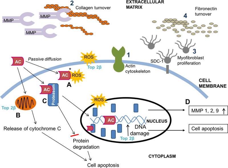
Nikitovic D, Juranek I, Wilks MF, Tzardi M, Tsatsakis A, Tzanakakis GN. Anthracycline-dependent cardiotoxicity and extracellular matrix remodeling. Chest 2014;146(4):1123–30. doi:10.1378/chest.14-0460.
*Outlines molecular basis for extracellular matrix remodeling.
-
-
- Curigliano G, Cardinale D, Suter T, Plataniotis G, de Azambuja E, Sandri MT et al. Cardiovascular toxicity induced by chemotherapy, targeted agents and radiotherapy: ESMO Clinical Practice Guidelines. Annals of oncology : official journal of the European Society for Medical Oncology / ESMO 2012;23 Suppl 7:vii155–66. doi:10.1093/annonc/mds293. - DOI - PubMed
Publication types
MeSH terms
Substances
Grants and funding
LinkOut - more resources
Full Text Sources
Other Literature Sources
Medical
Research Materials
