. 2016 Jun 30;166(1):102-14.
doi: 10.1016/j.cell.2016.05.032. Epub 2016 Jun 9.

Discovery of an Active RAG Transposon Illuminates the Origins of V(D)J Recombination

Affiliations

Discovery of an Active RAG Transposon Illuminates the Origins of V(D)J Recombination

Shengfeng Huang et al. Cell. .

Abstract

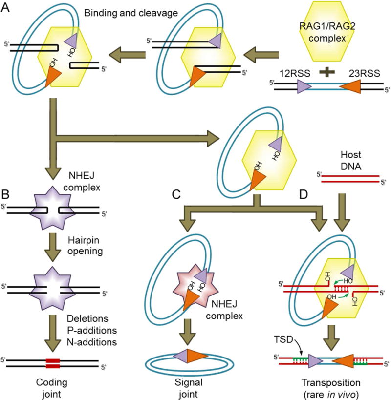
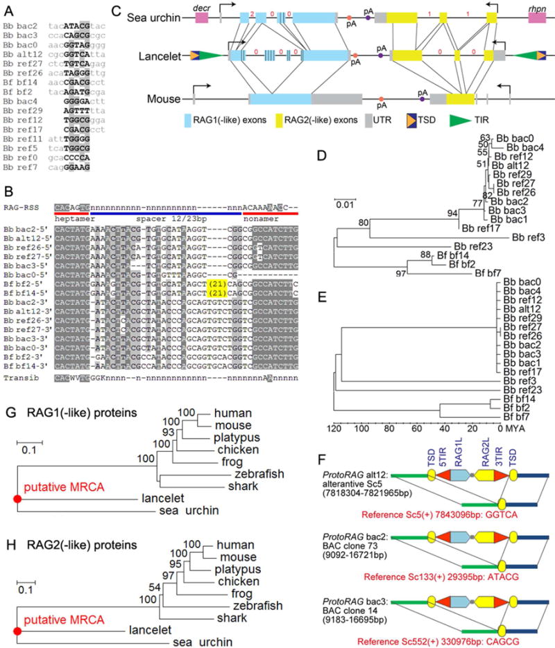
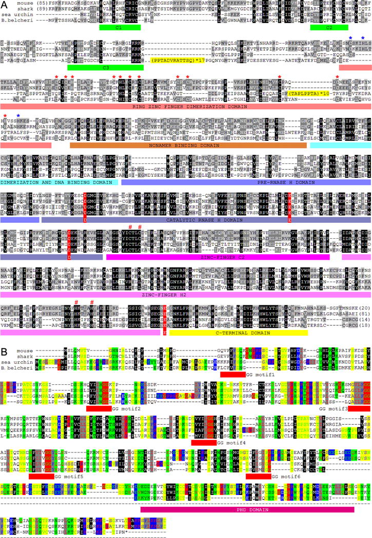
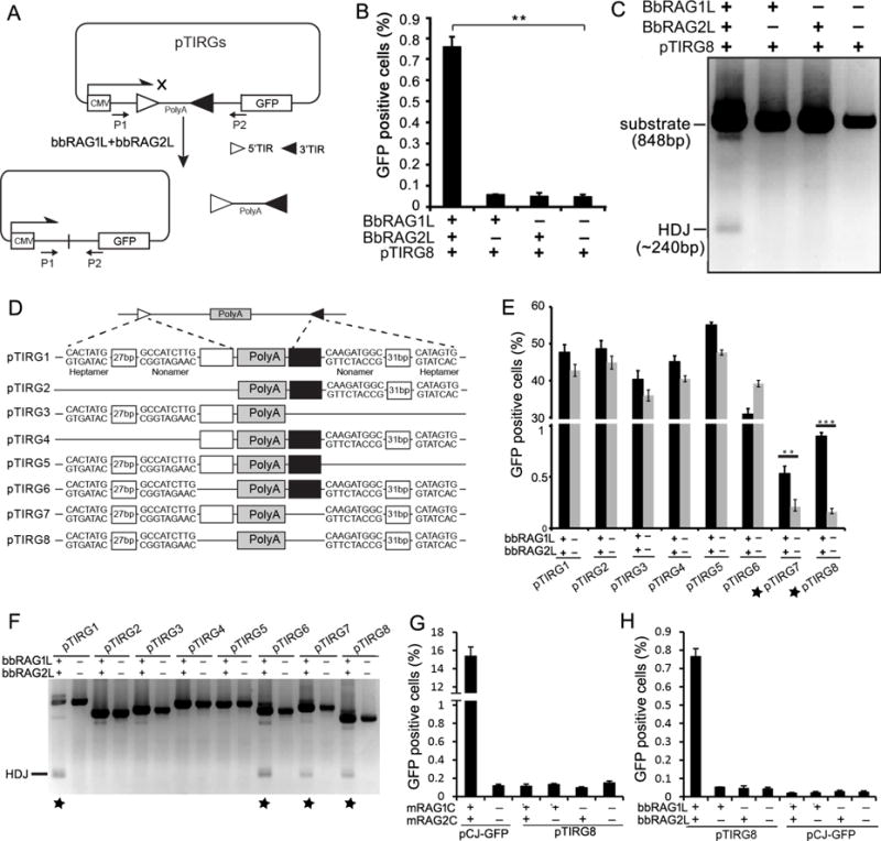
Co-option of RAG1 and RAG2 for antigen receptor gene assembly by V(D)J recombination was a crucial event in the evolution of jawed vertebrate adaptive immunity. RAG1/2 are proposed to have arisen from a transposable element, but definitive evidence for this is lacking. Here, we report the discovery of ProtoRAG, a DNA transposon family from lancelets, the most basal extant chordates. A typical ProtoRAG is flanked by 5-bp target site duplications and a pair of terminal inverted repeats (TIRs) resembling V(D)J recombination signal sequences. Between the TIRs reside tail-to-tail-oriented, intron-containing RAG1-like and RAG2-like genes. We demonstrate that ProtoRAG was recently active in the lancelet germline and that the lancelet RAG1/2-like proteins can mediate TIR-dependent transposon excision, host DNA recombination, transposition, and low-efficiency TIR rejoining using reaction mechanisms similar to those used by vertebrate RAGs. We propose that ProtoRAG represents a molecular "living fossil" of the long-sought RAG transposon.


Figures

Figure 1
Figure 1. Schematic of V(D)J recombination and transposition
(A) V(D)J recombination is initiated when the RAG complex binds a 12RSS/23RSS pair and cleaves the DNA, generating hairpin sealed coding ends and blunt RSS ends with a 3′ hydroxyl (OH) group. (B) The coding ends are nicked open by NHEJ DNA repair factors and then processed and joined, resulting in imprecise coding joints that can contain added nucleotides (red bars). (C) The cleaved RSS ends are thought to be bound initially by RAG, and subsequently are ligated together precisely by NHEJ repair factors to form a signal joint. (D) An alternative fate for the cleaved RSS ends bound to RAG is staggered attack by the 3′OH groups on a target DNA duplex (host DNA) resulting in insertion of the cleaved RSS fragment into the target and the creation of a flanking target site duplication (TSD). For RAG, this transposition outcome is rare in vivo but efficient in vitro.
Figure 2
Figure 2. Genomic sequence features of ProtoRAG
(A) Alignment of ProtoRAG TSDs and flanking sequences from the lancelet genome. (B) Alignment of ProtoRAG TIR sequences with the consensus RSS and Transib TIR. IUPAC codes used in the alignment: N=A, C, G or T; K=G or T; W=A, T; V=A, C or G. Lower case n indicates an undetermined nucleotide. Shading indicates sequence conservation, with darker gray indicating a higher degree of conservation. Bb: B. belcheri; Bf: B. floridae. ProtoRAG copy identification numbers correspond to those listed in Table S1. (C) Genomic organization of the lancelet ProtoRAG copy on BAC plasmid clone 73, sea urchin RAG1/2-like gene locus, and mouse RAG locus. Corresponding coding regions are indicated by thin lines. The phases of introns in coding regions are shown by red numbers. Repetitive regions in lancelet RAG1-like and sea urchin RAG1-like are marked by vertical bars. Terminal exons of flanking genes (decr, dienoyl-CoA reductase; rhpn, rhophilin) for the sea urchin RAG1/2-like locus are shown as purple boxes. (D) Neighbor-Joining trees of lancelet ProtoRAG copies assembled using Mega v5.2 (see Supplementary Methods). (E) Molecular dating analysis of lancelet ProtoRAG copies. This linearized tree with clock calibration was calculated using Mega v5.2. The root, or the divergence between B. belcheri and B. floridae, was set to 120 million years ago. (F) Three unfixed (polymorphic) ProtoRAG transposition events identified in B. belcheri genomes. Red text provides the coordinates of target sites and the sequence of TSDs on the reference genome. See Table S1 and Data S1 for further details. (G, H) Neighbor-joining trees of RAG1 (G) and RAG2 (H) protein homologs. MCRA, most common recent ancestor. See also Data S1 and Table S1.
Figure 3
Figure 3. The features of the proteins encoded by ProtoRAG
(A) Protein alignment of lancelet RAG1L with mouse RAG1, shark RAG1 and sea urchin RAG1L. Repeat motifs are highlighted in yellow. Three regions of conserved cysteine and histidine residues that might bind zinc are underlined with green bars. The N-terminal zinc binding dimerization domain is underlined with dark-red bars. The subdomains of the RAG1 core region are indicated with colored bars and labeled according to (Kim et al., 2015). The four conserved zinc ligand residues that contribute to proper folding of the RAG1 catalytic region are labeled with a red “#” (C727, C730, H937, H942 on mouse RAG1). The conserved acidic catalytic residues are highlighted with red shading (D600, E662, D708 and E962 on mouse RAG1). In the N-terminal RING-zinc finger dimerization domain (ZDD), fifteen zinc-coordinating residues (C266, H270, C290, C293, H295, C305, H307, C310, C313, C325, C328, C355, C360, H372 and H376 on mouse RAG1) that are conserved in vertebrate RAG1s are labeled with asterisks, with red asterisks indicating residues conserved in both vertebrate RAG1s and bbRAG1L, and blue asterisks indicating residues conserved in vertebrate RAG1s but not in lancelet RAG1L. GenBank accessions for mouse RAG1, shark RAG1, lancelet RAG2L and sea urchin RAG1L are NP_033045, XP_007886047, KJ748699 and NP_001028179, respectively. (B) Protein alignment of lancelet RAG2L with mouse RAG2, shark RAG2 and sea urchin RAG2L. Color shading shows the conservation of physiochemical properties. The N-terminal amino acid sequence can be grouped into Kelch-like repeats similar to Callebaut and Mornon (Callebaut and Mornon, 1998) and Fugmann et al (Fugmann et al., 2006). The central conserved GG motifs of the six Kelch-like repeats are underlined in red. The plant homeodomain (PHD) is also underlined below the alignment. GenBank accessions for mouse RAG2, shark RAG2, lancelet RAG2L and sea urchin RAG2L are NP_033046, XP_007885835, KJ748699 and NP_001028184, respectively. See also Figure S2.
Figure 4
Figure 4. TIR-dependent transposon excision mediated by bbRAG1L/2L ex vivo
(A) Diagram of the cell-based fluorescent reporter and PCR assay for DNA excision and recombination. Filled and unfilled triangles, 5′- and 3′-TIR sequences of ProtoRAG, respectively; P1/P2, PCR primers; CMV: cytomegalovirus promoter; PolyA: polyadenylation signal sequence. (B) Quantification of GFP positive cells by flow cytometry after transfection of 293T cells with pTIRG8 (containing the minimal ProtoRAG TIRs) with bbRAG1L and bbRAG2L expression vectors, as indicated. (C) PCR detection of HDJs from transfections of pTIRG8 as in (B). (D) Diagram of truncated TIR-containing substrates. Unfilled and filled boxes indicate the remainder of the 5′- and 3′-TIRs, respectively. (E) Quantification of GFP positive cells by flow cytometry after transfection of 293T cells with truncated TIR-containing substrates. (F) PCR detection of HDJs for truncated TIR-containing substrates. (G) Quantitation of GFP positive cells by flow cytometry after transfection of 293T cells with mouse RAG1 core and RAG2 core expression vectors with pTIRG8 (containing the minimal ProtoRAG TIRs) or pCJ-GFP (containing RSSs), as indicated. (H) Quantitation of GFP positive cells by flow cytometry after transfection of 293T cells with bbRAG1L and bbRAG2L expression vectors with pTIRG8 or pCJ-GFP (which contains a 12/23 RSS pair instead of the TIRs of pTIRG8), as indicated. See also Figure S3 and S4.
Figure 5
Figure 5. Biochemical analysis of DNA cleavage by bbRAG1L/2L in vitro
(A) Co-expressed, single-step purified bbRAG1L/2L and RAG1/2 proteins. *: background protein that elutes from amylose columns. (B) DNA cleavage substrates, with expected sizes of cleavage products indicated. (C) Time course of cleavage by bbRAG1L/2L (left) and RAG (right) as assessed by native polyacrylamide gel electrophoresis. *: prominent product corresponding to single cleavage at the 3′-TIR; **: central fragment resulting from double cleavage. All reactions contain HMGB1 and Mg2+ unless otherwise indicated. (D) Cleavage of TIR substrate (lanes 1–5) or RSS substrate (lanes 6–8) by the indicated proteins. D701A, bbRAG1L containing a D701A mutation combined with bbRAG2L. Lane 8, RAG1 core and RAG2 core proteins. (E) Cleavage by bbRAG1L/2L with different divalent cations (lanes 3–5) and in Mg2+ but in the absence of HMGB1 (lane 2). (F) Diagram of nick-hairpin mechanism of DNA cleavage. (G) Nicking and hairpinnning by bbRAG1L/2L as assessed by denaturing polyacrylamide gel electrophoresis. 3′-TIR DNA substrates, with 16 bp flanking the TIR on each side, were fluorescently 5′ end labeled on the top strand (filled circle) and were either intact (lanes 3–5) or pre-nicked immediately adjacent to the TIR (lanes 1–2). MUT: 3′-TIR substrate with a scrambled heptamer. M1 and M2: 16 nt and 32 nt markers. N: nick; HP: hairpin product. The hairpin product runs slightly faster than the 32 nt marker, likely due to a propensity to partially reanneal.
Figure 6
Figure 6. Ex vivo analysis of the host DNA rejoining and transposon self-resealing after transposon excision by bbRAG1L/2L
(A) The bacterial colony assay and the plasmid (pTIR104) designed to detect bbRAG1L/bbRAG2L mediated transposon excision and HDJ rejoining. The results are shown in Figure S5. (B) The bacterial colony assay designed to detect bbRAG1L/bbRAG2L mediated complex transposon self-resealing events after transposon excision. The resulted recombinants are categorized by the junctions identified by DNA sequencing. The left panel shows the plasmid (pTIR204) designed for the detection, in which the TIRs are retained with the backbone of the plasmid after cleavage. See also Figure S5.
Figure 7
Figure 7. In vitro and ex vivo intermolecular transposition mediated by bbRAG1L/2L
(A) Schematic diagram of the assay used to detect in vitro transposition mediated by purified bbRAG1L and bbRAG2L proteins. (B) The distribution of in vitro transposition target sites in the recipient plasmid. (C) Quantitation of in vitro transposition efficiency of bbRAG1L/2L and mouse RAG1/2. Each dot represents the results of an independent reaction with the horizontal bar indicating the mean (+/− SEM). For RAG1/2, one data point (red) was outside of the range of the y-axis and its value is indicated in parentheses. Means for bbRAG1L/2L and RAG1/2 were 19.4 and 44.6, respectively. (D) Schematic diagram of the assay used to detect in vivo transposition mediated by bbRAG1L and bbRAG2L in 293T cells. (E) The distribution of in vivo transposition target sites in the recipient plasmid. See also Figure S6.

Comment in

Similar articles

Cited by

References

    1. Agrawal A, Eastman QM, Schatz DG. Transposition mediated by RAG1 and RAG2 and its implications for the evolution of the immune system. Nature. 1998;394:744–751. - PubMed
    1. Callebaut I, Mornon JP. The V(D)J recombination activating protein RAG2 consists of a six-bladed propeller and a PHD fingerlike domain, as revealed by sequence analysis. Cell Mol Life Sci. 1998;54:880–891. - PMC - PubMed
    1. Chatterji M, Tsai CL, Schatz DG. Mobilization of RAG-generated signal ends by transposition and insertion in vivo. Mol Cell Biol. 2006;26:1558–1568. - PMC - PubMed
    1. Corneo B, Benmerah A, Villartay JP. A short peptide at the C terminus is responsible for the nuclear localization of RAG2. Eur J Immunol. 2002;32:2068–2073. - PubMed
    1. Corneo B, Wendland RL, Deriano L, Cui X, Klein IA, Wong SY, Arnal S, Holub AJ, Weller GR, Pancake BA, et al. Rag mutations reveal robust alternative end joining. Nature. 2007;449:483–486. - PubMed

Publication types