

CETP Lowers TLR4 Expression Which Attenuates the Inflammatory Response Induced by LPS and Polymicrobial Sepsis
- PMID: 27293313
- PMCID: PMC4880711
- DOI: 10.1155/2016/1784014
CETP Lowers TLR4 Expression Which Attenuates the Inflammatory Response Induced by LPS and Polymicrobial Sepsis
Abstract
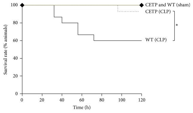
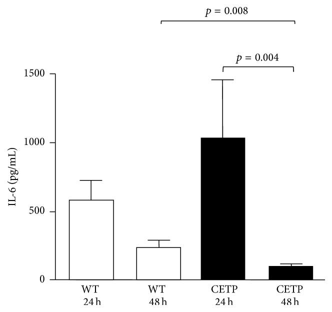
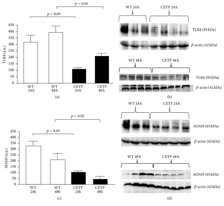
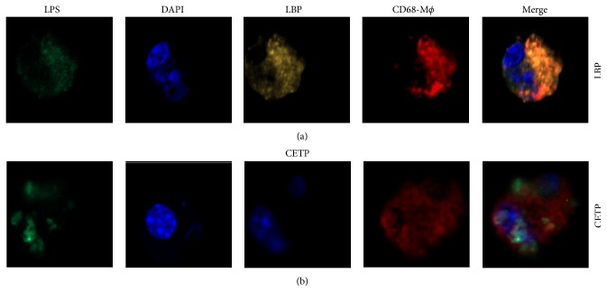
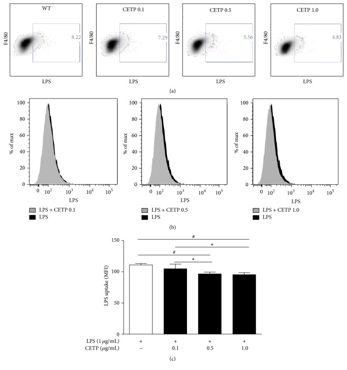
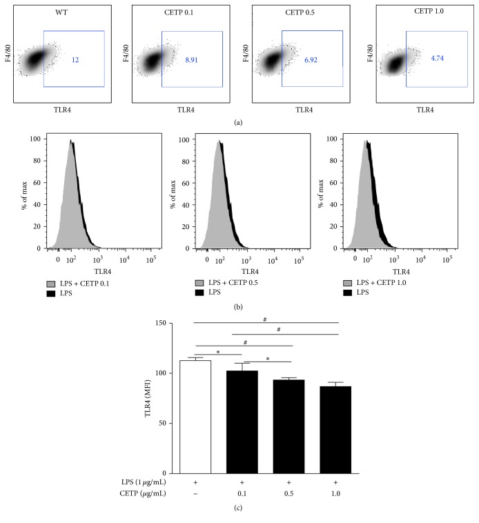
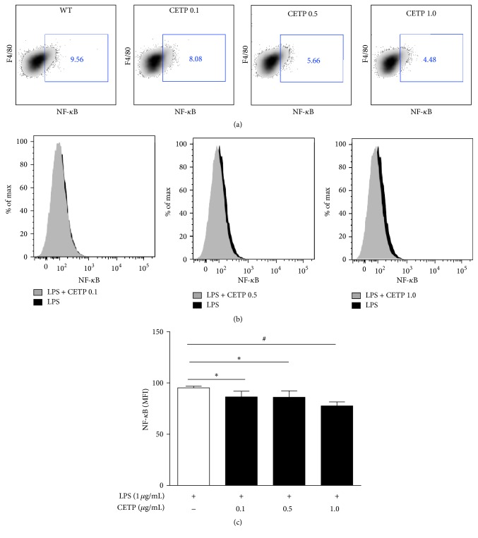
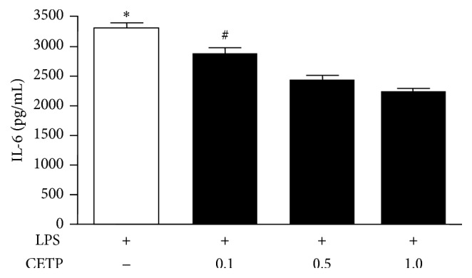
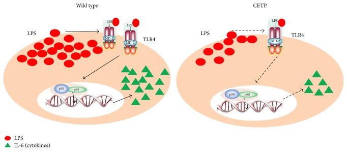
Sepsis is a systemic inflammatory response to infection eliciting high mortality rate which is a serious health problem. Despite numerous studies seeking for therapeutic alternatives, the mechanisms involved in this disease remain elusive. In this study we evaluated the influence of cholesteryl ester transfer protein (CETP), a glycoprotein that promotes the transfer of lipids between lipoproteins, on the inflammatory response in mice. Human CETP transgenic mice were compared to control mice (wild type, WT) after polymicrobial sepsis induced by cecal ligation and puncture (CLP), aiming at investigating their survival rate and inflammatory profiles. Macrophages from the peritoneal cavity were stimulated with LPS in the presence or absence of recombinant CETP for phenotypic and functional studies. In comparison to WT mice, CETP mice showed higher survival rate, lower IL-6 plasma concentration, and decreased liver toll-like receptor 4 (TLR4) and acyloxyacyl hydrolase (AOAH) protein. Moreover, macrophages from WT mice to which recombinant human CETP was added decreased LPS uptake, TLR4 expression, NF-κB activation and IL-6 secretion. This raises the possibility for new therapeutic tools in sepsis while suggesting that lowering CETP by pharmacological inhibitors should be inconvenient in the context of sepsis and infectious diseases.
Figures
Similar articles
-
Human cholesteryl ester transfer protein lacks lipopolysaccharide transfer activity, but worsens inflammation and sepsis outcomes in mice.J Lipid Res. 2021;62:100011. doi: 10.1194/jlr.RA120000704. Epub 2020 Dec 15. J Lipid Res. 2021. PMID: 33500240 Free PMC article.
-
Kallistatin treatment attenuates lethality and organ injury in mouse models of established sepsis.Crit Care. 2015 May 1;19(1):200. doi: 10.1186/s13054-015-0919-4. Crit Care. 2015. PMID: 25930108 Free PMC article.
-
Anti-inflammatory effects of benzenediamine derivate FC-98 on sepsis injury in mice via suppression of JNK, NF-κB and IRF3 signaling pathways.Mol Immunol. 2015 Oct;67(2 Pt B):183-92. doi: 10.1016/j.molimm.2015.05.005. Epub 2015 May 29. Mol Immunol. 2015. PMID: 26032013
-
Biochemical transformation of bacterial lipopolysaccharides by acyloxyacyl hydrolase reduces host injury and promotes recovery.J Biol Chem. 2020 Dec 18;295(51):17842-17851. doi: 10.1074/jbc.REV120.015254. J Biol Chem. 2020. PMID: 33454018 Free PMC article. Review.
-
New insights into lipopolysaccharide inactivation mechanisms in sepsis.Biomed Pharmacother. 2021 Sep;141:111890. doi: 10.1016/j.biopha.2021.111890. Epub 2021 Jul 3. Biomed Pharmacother. 2021. PMID: 34229252 Review.
Cited by
-
The impact of cholesteryl ester transfer protein on the progression of cutaneous leishmaniasis.Front Immunol. 2024 Jun 20;15:1389551. doi: 10.3389/fimmu.2024.1389551. eCollection 2024. Front Immunol. 2024. PMID: 38966642 Free PMC article.
-
Impact of High-Density Lipoproteins on Sepsis.Int J Mol Sci. 2022 Oct 26;23(21):12965. doi: 10.3390/ijms232112965. Int J Mol Sci. 2022. PMID: 36361756 Free PMC article. Review.
-
Cholesteryl ester transfer protein (CETP), HDL capacity of receiving cholesterol and status of inflammatory cytokines in patients with severe heart failure.Lipids Health Dis. 2018 Oct 20;17(1):242. doi: 10.1186/s12944-018-0888-0. Lipids Health Dis. 2018. PMID: 30342531 Free PMC article.
-
Effects of Anti-IL-17 on Inflammation, Remodeling, and Oxidative Stress in an Experimental Model of Asthma Exacerbated by LPS.Front Immunol. 2018 Jan 5;8:1835. doi: 10.3389/fimmu.2017.01835. eCollection 2017. Front Immunol. 2018. PMID: 29379497 Free PMC article.
-
Stratification of Sepsis Patients on Admission into the Intensive Care Unit According to Differential Plasma Metabolic Phenotypes.J Proteome Res. 2024 Apr 5;23(4):1328-1340. doi: 10.1021/acs.jproteome.3c00803. Epub 2024 Mar 21. J Proteome Res. 2024. PMID: 38513133 Free PMC article.
References
Publication types
MeSH terms
Substances
LinkOut - more resources
Full Text Sources
Other Literature Sources
Medical
Miscellaneous
