

Early retinal inflammatory biomarkers in the middle cerebral artery occlusion model of ischemic stroke
- PMID: 27293375
- PMCID: PMC4893077
Early retinal inflammatory biomarkers in the middle cerebral artery occlusion model of ischemic stroke
Abstract
Purpose: The transient middle cerebral artery occlusion (MCAO) model of stroke is one of the most commonly used models to study focal cerebral ischemia. This procedure also results in the simultaneous occlusion of the ophthalmic artery that supplies the retina. Retinal cell death is seen days after reperfusion and leads to functional deficits; however, the mechanism responsible for this injury has not been investigated. Given that the eye may have a unique ocular immune response to an ischemic challenge, this study examined the inflammatory response to retinal ischemia in the MCAO model.
Methods: Young male C57B/6 mice were subjected to 90-min transient MCAO and were euthanized at several time points up to 7 days. Transcription of inflammatory cytokines was measured with quantitative real-time PCR, and immune cell activation (e.g., phagocytosis) and migration were assessed with ophthalmoscopy and flow cytometry.
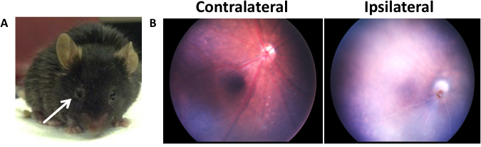
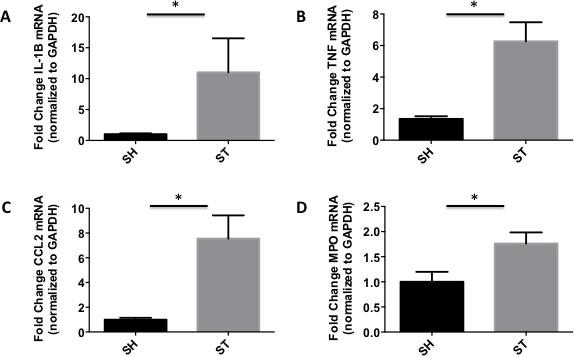
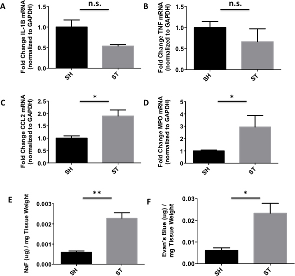
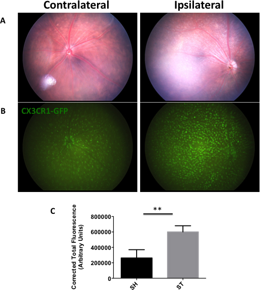
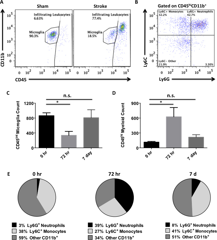
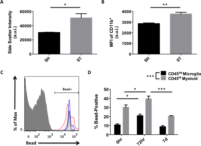
Results: Observation of the affected eye revealed symptoms consistent with Horner's syndrome. Light ophthalmoscopy confirmed the reduced blood flow of the retinal arteries during occlusion. CX3CR1-GFP reporter mice were then employed to evaluate the extent of the ocular microglia and monocyte activation. A significant increase in green fluorescent protein (GFP)-positive macrophages was seen throughout the ischemic area compared to the sham and contralateral control eyes. RT-PCR revealed enhanced expression of the monocyte chemotactic molecule CCL2 early after reperfusion followed by a delayed increase in the proinflammatory cytokine TNF-α. Further analysis of peripheral leukocyte recruitment by flow cytometry determined that monocytes and neutrophils were the predominant immune cells to infiltrate at 72 h. A transient reduction in retinal microglia numbers was also observed, demonstrating the ischemic sensitivity of these cells. Blood-eye barrier permeability to small and large tracer molecules was increased by 72 h. Retinal microglia exhibited enhanced phagocytic activity following MCAO; however, infiltrating myeloid cells were significantly more efficient at phagocytizing material at all time points. Immune homeostasis in the affected eye was largely restored by 7 days.
Conclusions: This work demonstrates that there is a robust inflammatory response in the eye following MCAO, which may contribute to a worsening of retinal injury and visual impairment. These results mirror what has been observed in the brain after MCAO, suggesting a conserved inflammatory signaling response to ischemia in the central nervous system. Imaging of the eye may therefore serve as a useful non-invasive prognostic indicator of brain injury after MCAO. Future studies are needed to determine whether this inflammatory response is a potential target for therapeutic manipulation in retinal ischemia.
Figures
Similar articles
-
miR-221 Exerts Neuroprotective Effects in Ischemic Stroke by Inhibiting the Proinflammatory Response.J Stroke Cerebrovasc Dis. 2021 Feb;30(2):105489. doi: 10.1016/j.jstrokecerebrovasdis.2020.105489. Epub 2020 Dec 1. J Stroke Cerebrovasc Dis. 2021. PMID: 33276305
-
Functional differences between microglia and monocytes after ischemic stroke.J Neuroinflammation. 2015 May 29;12:106. doi: 10.1186/s12974-015-0329-1. J Neuroinflammation. 2015. PMID: 26022493 Free PMC article.
-
Interleukin-19 alleviates brain injury by anti-inflammatory effects in a mice model of focal cerebral ischemia.Brain Res. 2016 Nov 1;1650:172-177. doi: 10.1016/j.brainres.2016.09.006. Epub 2016 Sep 5. Brain Res. 2016. PMID: 27608956
-
Modeling Transient Focal Ischemic Stroke in Rodents by Intraluminal Filament Method of Middle Cerebral Artery Occlusion.Methods Mol Biol. 2018;1717:101-113. doi: 10.1007/978-1-4939-7526-6_9. Methods Mol Biol. 2018. PMID: 29468587 Review.
-
Inflammation as a therapeutic target in acute ischemic stroke treatment.Curr Top Med Chem. 2009;9(14):1240-60. doi: 10.2174/156802609789869619. Curr Top Med Chem. 2009. PMID: 19849665 Review.
Cited by
-
Eye Opener in Stroke.Stroke. 2019 Aug;50(8):2197-2206. doi: 10.1161/STROKEAHA.119.025249. Epub 2019 Jun 27. Stroke. 2019. PMID: 31242827 Free PMC article.
-
Transfer of Mitochondria from Healthy Stem Cells to Injured Cells in Stroke with Retinal Impairments.Methods Mol Biol. 2025;2960:125-138. doi: 10.1007/7651_2024_599. Methods Mol Biol. 2025. PMID: 39930314
-
Short-Time Ocular Ischemia Induces Vascular Endothelial Dysfunction and Ganglion Cell Loss in the Pig Retina.Int J Mol Sci. 2019 Sep 21;20(19):4685. doi: 10.3390/ijms20194685. Int J Mol Sci. 2019. PMID: 31546635 Free PMC article.
-
Morphological and functional observations of a novel model of retinal ischemia injury induced by bilateral carotid artery stenosis in mice.Int J Ophthalmol. 2024 Dec 18;17(12):2192-2202. doi: 10.18240/ijo.2024.12.06. eCollection 2024. Int J Ophthalmol. 2024. PMID: 39697883 Free PMC article.
-
The temporal dynamic of bradykinin type 2 receptor effects reveals its neuroprotective role in the chronic phase of cerebral and retinal ischemic injury.J Cereb Blood Flow Metab. 2025 Jan;45(1):153-170. doi: 10.1177/0271678X241270241. Epub 2024 Aug 7. J Cereb Blood Flow Metab. 2025. PMID: 39113417 Free PMC article.
References
-
- Liu F, McCullough LD. Middle cerebral artery occlusion model in rodents: methods and potential pitfalls. J Biomed Biotechnol. 2011;2011:464701. http://www.ncbi.nlm.nih.gov/entrez/query.fcgi?cmd=Retrieve&db=PubMed&lis... - PMC - PubMed
-
- Mohr JP. Some clinical aspects of acute stroke. Excellence in Clinical Stroke Award Lecture. Stroke. 1997;28:1835–9. http://www.ncbi.nlm.nih.gov/entrez/query.fcgi?cmd=Retrieve&db=PubMed&lis... - PubMed
-
- Ng YS, Stein J, Ning M, Black-Schaffer RM. Comparison of clinical characteristics and functional outcomes of ischemic stroke in different vascular territories. Stroke. 2007;38:2309–14. http://www.ncbi.nlm.nih.gov/entrez/query.fcgi?cmd=Retrieve&db=PubMed&lis... - PubMed
-
- Steele EC, Jr, Guo Q, Namura S. Filamentous middle cerebral artery occlusion causes ischemic damage to the retina in mice. Stroke. 2008;39:2099–104. http://www.ncbi.nlm.nih.gov/entrez/query.fcgi?cmd=Retrieve&db=PubMed&lis... - PubMed
-
- Shuaib A, Butcher K, Mohammad AA, Saqqur M, Liebeskind DS. Collateral blood vessels in acute ischaemic stroke: a potential therapeutic target. Lancet Neurol. 2011;10:909–21. http://www.ncbi.nlm.nih.gov/entrez/query.fcgi?cmd=Retrieve&db=PubMed&lis... - PubMed
Publication types
MeSH terms
Substances
Grants and funding
LinkOut - more resources
Full Text Sources
Medical
