. 2016 Jan 20;26(3):421-436.
doi: 10.1002/adfm.201504160. Epub 2015 Dec 8.

Silk Biomaterials with Vascularization Capacity

Affiliations

Silk Biomaterials with Vascularization Capacity

Hongyan Han et al. Adv Funct Mater. .

Abstract

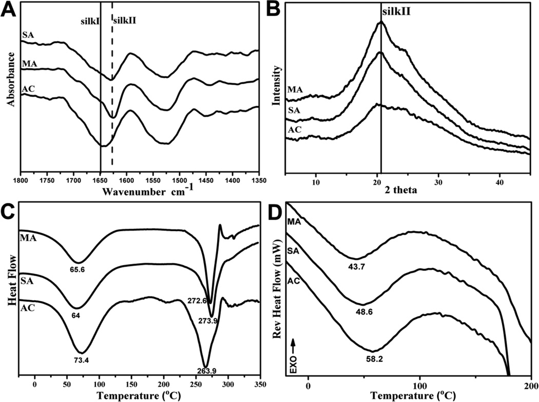
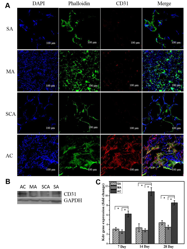
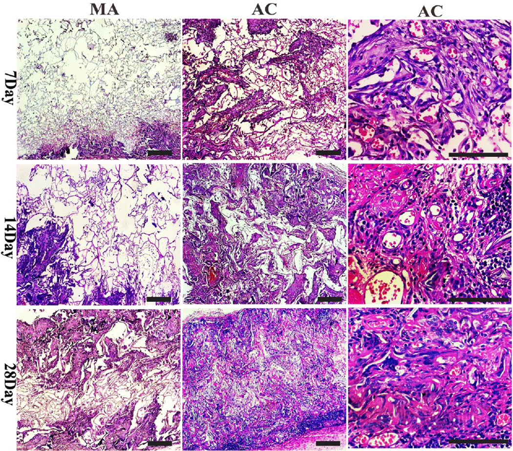
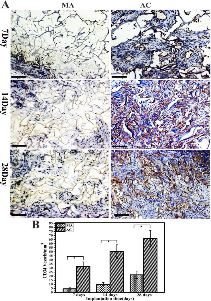
Functional vascularization is critical for the clinical regeneration of complex tissues such as kidney, liver or bone. The immobilization or delivery of growth factors has been explored to improve vascularization capacity of tissue engineered constructs, however, the use of growth factors has inherent problems such as the loss of signaling capability and the risk of complications such as immunological responses and cancer. Here, a new method of preparing water-insoluble silk protein scaffolds with vascularization capacity using an all aqueous process is reported. Acid was added temporally to tune the self-assembly of silk in lyophilization process, resulting in water insoluble scaffold formation directly. These biomaterials are mainly noncrystalline, offering improved cell proliferation than previously reported silk materials. These systems also have appropriate softer mechanical property that could provide physical cues to promote cell differentiation into endothelial cells, and enhance neovascularization and tissue ingrowth in vivo without the addition of growth factors. Therefore, silk-based degradable scaffolds represent an exciting biomaterial option, with vascularization capacity for soft tissue engineering and regenerative medicine.

Keywords: lyophilization; silk; soft tissue engineering; stiffness; vascularization.


Figures

Figure 1
Figure 1
SEM morphologies of silk scaffolds prepared through different processes: The samples were as follows: SA, silk scaffolds prepared by salt-leaching process; MA, Methanol-treated silk scaffolds derived from silk solution with 6.7% silk nanofiber; AC, Acid-assisted silk scaffolds derived from silk solution with 6.7% silk nanofiber. The right images showed the nanoscaled topography of pore wall with high magnification.
Figure 2
Figure 2
FTIR (A), XRD (B), DSC (C) and TMDSC (D) characterization of silk scaffolds prepared through different processes: The samples were as follows: SA, silk scaffolds prepared by salt-leaching process; MA, Methanol-treated silk scaffolds derived from silk solution with 6.7% silk nanofiber; AC, Acid-assisted silk scaffolds derived from silk solution with 6.7% silk nanofiber.
Figure 3
Figure 3
Enzyme degradation (A) and mechanical properties (B, C) of silk scaffolds prepared through different processes. The samples were as follows: SA, silk scaffolds prepared by salt-leaching process; MA, Methanol-treated silk scaffolds derived from silk solution with 6.7% silk nanofiber; AC, Acid-assisted silk scaffolds derived from silk solution with 6.7% silk nanofiber. In enzyme degradation process, the samples were cultured in protease XIV solution (5 U ml−1 in PBS) at 37°C. Typical stress-strain curves (B) and compressive modulus (C) of the scaffolds were measured in wet conditions.
Figure 4
Figure 4
The proliferation behavior of BMSCs on different silk scaffolds: (A) Fluorescence microscopy images of BMSC cultured on the samples at day 12, Blue (DAPI) for nuclei and silk fibroin scaffolds; green (FITC labeled phalloidin) for F-actin; (B) SEM images of BMSCs on the samples at day 12; (C) BMSC proliferation on the samples measured with DNA analysis. *statistically significant P<0.05. The samples were as follows: SA, silk scaffolds prepared by salt-leaching process; MA, Methanol-treated silk scaffolds derived from silk solution with 6.7% silk nanofiber; AC, Acid-assisted silk scaffolds derived from silk solution with 6.7% silk nanofiber.
Figure 5
Figure 5
BMSC differentiation into endothelial cells on different silk scaffolds: (A) the expression of CD31 (red) in different scaffolds at day 28 by confocal microscopy, blue (DAPI) for nuclei and silk fibroin scaffolds, green (FITC labeled phalloidin) for F-actin; (B) the expression of CD31 at day 28 detected by western blot; (C) mRNA levels of KDR quantified by real-time PCR. The samples were as follows: SA, silk scaffolds prepared by salt-leaching process; MA, methanol-treated silk scaffolds derived from silk solution with 6.7% silk nanofiber; SAC, silk scaffolds prepared by an acid-assisted salt-leaching process; AC, acid-assisted silk scaffolds derived from silk solution with 6.7% silk nanofiber. The expression of KDR is normalized to GAPDH. * statistically significant P<0.05.
Figure 6
Figure 6
HE staining images of sections of AC and MA implants at 7, 14 and 28 days after implantation in vivo. Representative section images revealed better neovascularization and tissue ingrowth in AC implant. The right and middle pictures were the low magnification morphology of MA and AC samples (scale bar = 200 µm), while the right pictures showed the AC morphology at high magnification (scale bar = 100 µm). Protoplasm stained with picrosirius red and nuclei stained with blue.
Figure 7
Figure 7
(A) Immunohistochemistry staining images of sections of AC and MA implants at 7, 14 and 28 days after implantation in vivo (scale bar = 200µm). The microvessels were stained with dark gray positive for CD34, and nuclei was stained with blue; (B) Vessel density within the AC and MA scaffolds after implantation into the lateral incisions on the dorsal region. * statistically significant P<0.05
Figure 8
Figure 8
Masson trichrome staining images of sections of AC and MA implants at 7, 14 and 28 days after implantation in vivo. Collagen fibers are stained blue, nuclei are stained dark red/purple, and cytoplasm is stained pink.

References

    1. Stahl PJ, Chan TR, Shen YI, Sun G, Gerecht S, Yu SM. Adv. Funct. Mater. 2014;24:3213. - PMC - PubMed
    1. Mansbridge J. J. Biomater. Sci. 2008;19:955. - PubMed
    1. Temenoff JS, Mikos AG. Biomaterials. 2000;21:431. - PubMed
    1. Wray LS, Tsioris K, Gil ES, Omenetto FG, Kaplan DL. Adv. Funct. Mater. 2013;23:3404. - PMC - PubMed
    1. Griffith LG, Naughton G. Science. 2002;295:1009. - PubMed