

Fibrous Dysplasia with Massive Cartilaginous Differentiation (Fibrocartilaginous Dysplasia) in the Proximal Femur: A Case Report and Review of the Literature
- PMID: 27293399
- PMCID: PMC4899633
- DOI: 10.1159/000443476
Fibrous Dysplasia with Massive Cartilaginous Differentiation (Fibrocartilaginous Dysplasia) in the Proximal Femur: A Case Report and Review of the Literature
Abstract
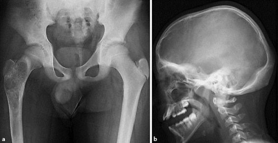
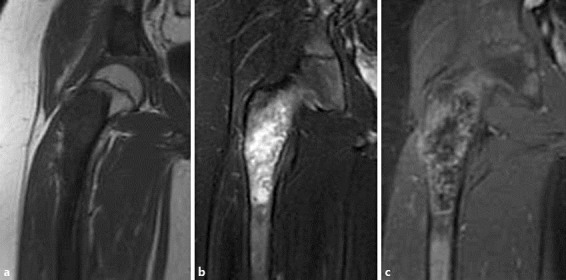
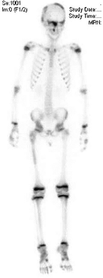
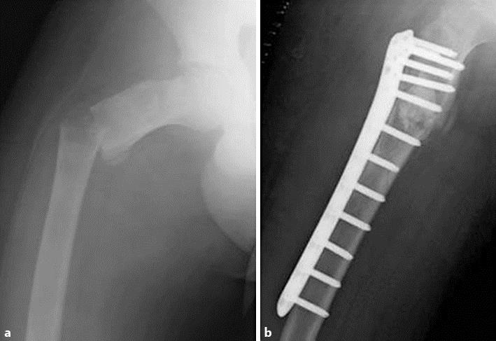
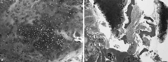
Fibrous dysplasia (FD) is a monostotic or polyostotic benign bone lesion with spindle-cell proliferation in woven bone and stroma. Rarely, cartilaginous differentiation can be seen in the lesions of FD. FD with massive cartilaginous differentiation is called fibrocartilaginous dysplasia (FCD) and is considered a rare variant of FD. Although pathological findings of FD show irregular immature bone formation without osteoblastic rimming in fibrous tissue, and rarely show very small amounts of cartilage, histological images of FCD are said to show that cartilage with a relatively high cell density is present in the majority and that FD-like findings are seen in parts of it. The most characteristic feature of FCD on images is calcification in the lesions reflecting cartilaginous tissue. On the other hand, typical radiographic findings of FD include shadows with a ground-glass appearance and thinning and bulging of the cortical bone, the observation if calcification is not usual. Therefore, in the diagnosis of FCD, differentiation from multiple enchondromatosis, Ollier disease, chondrosarcoma, and chondrosarcoma secondary to FD is necessary, and it seems important to make a careful diagnosis based not only on the pathological findings but also on imaging and clinical findings. Herein, we report on a case of FD of the proximal femur associated with intralesional extensive carti laginous differentiation in which a pathological fracture occurred during follow-up, with a review of the literature.
Keywords: Cartilaginous differentiation; Fibrocartilaginous dysplasia; Fibrous dysplasia; Proximal femur.
Figures
References
-
- Dorfman HD, Czeniak B. Bone Tumors. St. Louis: Mosby; 1998.
-
- Tabareau-Delalande F, Collin C, Gomez-Brouchet A, Decouvelaere AV, Bouvier C, Larousserie F, Marie B, Delfour C, Aubert S, Rosset P, de Muret A, Pagés JC, de Pinieux G. Diagnostic value of investigating GNAS mutations in fibro-osseous lesions: a retrospective study of 91 cases of fibrous dysplasia and 40 other fibro-osseous lesions. Mod Pathol. 2013;26:911–921. - PubMed
-
- Unni KK, Inwards CY. Dahlin's Bone Tumors rev. ed 6. Philadelphia Lippincott: Williams and Wilkins; 2009.
-
- Muezzinoglu B, Oztop F. Fibrocartilaginous dysplasia: a variant of fibrous dysplasia. Malays J Pathol. 2001;23:35–39. - PubMed
-
- Pelzmann KS, Nagel DZ, Salyer WR. Case report 114. Skeletal Radiol. 1980;5:116–118. - PubMed
Publication types
LinkOut - more resources
Full Text Sources
Other Literature Sources
