Review
. 2016:2016:8304879.
doi: 10.1155/2016/8304879. Epub 2016 May 15.

Stem Cell Therapy for Treatment of Ocular Disorders

Affiliations
Review

Stem Cell Therapy for Treatment of Ocular Disorders

Padma Priya Sivan et al. Stem Cells Int. 2016.

Abstract

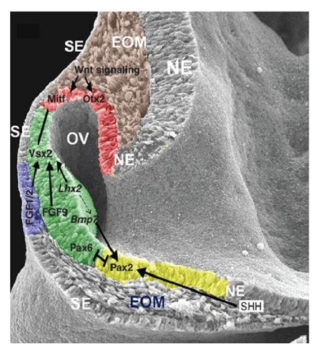
Sustenance of visual function is the ultimate focus of ophthalmologists. Failure of complete recovery of visual function and complications that follow conventional treatments have shifted search to a new form of therapy using stem cells. Stem cell progenitors play a major role in replenishing degenerated cells despite being present in low quantity and quiescence in our body. Unlike other tissues and cells, regeneration of new optic cells responsible for visual function is rarely observed. Understanding the transcription factors and genes responsible for optic cells development will assist scientists in formulating a strategy to activate and direct stem cells renewal and differentiation. We review the processes of human eye development and address the strategies that have been exploited in an effort to regain visual function in the preclinical and clinical state. The update of clinical findings of patients receiving stem cell treatment is also presented.


Figures

Figure 1
Figure 1
Crucial biomolecules expression in an embryonic mouse at 9.5 days. The neural ectoderm (NE) bulges as optic vesicle (OV) to reach the surface ectoderm (SE) on both sides. The SE became thicker upon the contact of NE to become the lens placode. Except in the lens placode region, the NE and SE are separated by the EOM. In the NE, the presumptive RPE, neural retina, and optic tract are colored red, green, and yellow, respectively. The lens placode is colored blue. The TF reciprocally act to regulate eye development. EOM, extraocular mesenchyme; RPE, retinal pigmented epithelium; NR, neural retina; TF, transcription factor. Copyright 2012. Modified with permission from Cold Spring Harbor Laboratory Press [26].
Figure 2
Figure 2
CMZ in vertebrates. The CMZ is progressively reduced in higher vertebrates. The adult eye of different vertebrates (frogs and fish (a), avians (b), and mammals (c)) is shown in blue and represents the neural retina of embryonic origin, which lacks the continuous renewal ability of the CMZ, which is shown in yellow. CMZ: ciliary marginal zone. Copyright 2004. Modified with permission from UBC Press [27].
Figure 3
Figure 3
Flow chart: major events of eye development and the involvement of biomolecules. Copyright 2009. Modified with permission from Mosby/Elsevier Ltd. [23].
Figure 4
Figure 4
Tracking of injected human Wharton's jelly-derived MSCs in an RP rat model (Royal College of Surgeons rats) with microcomputed tomography. Microcomputed tomography images show localization of gold-loaded human Wharton's jelly-derived MSCs in the right eye (a) on day one. The cells were found to be retained in the eye without further migration at day thirty (b) and day seventy (c) after transplantation. PKH 26 (labelled red) indicated the subretinal site of human Wharton's jelly-derived MSCs after cell transplantation at week two. Modified with permission from Creative Commons Attribution License [103].
Figure 5
Figure 5
Observation of differentiation of human Wharton's jelly-derived MSCs into retinal cell phenotypes in RCS rats by confocal microscopy. Confocal microscopy picture of the whole eye (A) and magnified pictures of the transplanted region (B–D). The red box indicates the magnified region, and the white arrow demonstrates the transplanted region. The antibodies used were anti-PKC-α (bipolar cell), anti-human/rat rhodopsin (rod photoreceptor), anti-human stem 121 (MSC), and anti-GFAP (Müller glial cells). DAPI was used to stain the nucleus in the retinal layer. Colocalization of DAPI (blue) and stem 121 (red) with PKC-α (green), GFAP (green), and rhodopsin (green) was found at day seventy after transplantation, suggesting that human Wharton's jelly-derived MSCs have the ability to differentiate into retinal neurons or to fuse with the degenerating neurons. Scale bar indicates 10 μm. Modified with permission from Creative Commons Attribution License [103].
Figure 6
Figure 6
Electron microscopy pictures of telocyte-putative stem cell junctions observed in the human heart. The picture describes the contact point (arrowheads) among a putative stem cell and a telocyte (blue color). Broader planar contacts (double arrows) can be observed. (a) The average distance between the putative stem cell and plasma membranes of telopode, Tp, is 43 ± 20.3 nm (min: 20.3 nm; max: 90.6 nm). E, endothelial cell; sv, shed vesicles; CM, cardiomyocyte. (b) High magnification on a consecutive ultrathin region of the rectangular site indicated in (a) describes the geometry of the 8 μm long heterocellular connections; plasma membranes of tight-fitting apposed sectors (double arrows); dot contacts (arrowheads) change with planar contacts. A small cellular projection of putative stem cell (arrow) is located on a small recess of the telocyte. Thick nanostructures (15–20 nm) can be found with connection of the plasma membranes of the cells (white arrowheads). Bars represent 2 μm. Modified with permission from Creative Commons Attribution License [116].
Figure 7
Figure 7
Pictures of eye fundus with pigmentation where retinal pigment epithelium differentiated from human ESCs was transplanted. (a–c) Color fundus pictures and images of spectral domain-optical coherence tomography at baseline of patient eyes of ARMD (dotted circle indicates an outline of the site of cell transplantation) and at an eye after 3 and 6 months of the transplantation. A pigmented patch of transplanted cells (arrows in (b) and (c)) grows bigger and has more pigmentation in six months. Optical coherence tomography (inset of figures) indicates the existence of cells on the inner sites of Bruch's membrane at six months compared with the baseline of the eye. (d–f) Color fundus pictures and pictures of spectral domain-optical coherence tomography at baseline of patient eyes of Stargardt's macular dystrophy (dotted circle indicates an outline of the site of cell transplantation) and an eye after six and twelve months after transplantation. Patches of transplanted cells exist around the edge of baseline atrophy in retinal pigment epithelium (e), which grow more significant after twelve months (arrows in (f)). Pictures of spectral domain-optical coherence tomography at baseline (d) and six months (e) indicate that the enhancement of pigmentation is found at the level of normal monolayer retinal pigment epithelium engraftment, the retinal pigment epithelium, and survival at six months (arrows in (e)), which is close to the site of bare Bruch's membrane being lack of native retinal pigment epithelium. (g–i) Color fundus pictures of a patient of Stargardt's macular dystrophy (dotted circle indicates an outline of the transplantation site). A big central site of atrophy can be seen on the preoperative picture (g). A site of transplantation of retinal pigment epithelium cells can be seen at the superior half of the atrophic lesion at six months (h), which grows bigger and has more pigmentation at fifteen months (i). Copyright 2015. Modified with permission from Elsevier Ltd. [72].

References

    1. Shichida Y., Matsuyama T. Evolution of opsins and phototransduction. Philosophical Transactions of the Royal Society B: Biological Sciences. 2009;364(1531):2881–2895. doi: 10.1098/rstb.2009.0051. - DOI - PMC - PubMed
    1. Jonas J. B., George R., Asokan R., et al. Prevalence and causes of vision loss in central and south Asia: 1990–2010. British Journal of Ophthalmology. 2014;98(5):592–598. doi: 10.1136/bjophthalmol-2013-303998. - DOI - PubMed
    1. Bourne R. R. A., Jonas J. B., Flaxman S. R., et al. Prevalence and causes of vision loss in high-income countries and in Eastern and Central Europe: 1990–2010. British Journal of Ophthalmology. 2014;98(5):629–638. doi: 10.1136/bjophthalmol-2013-304033. - DOI - PubMed
    1. Vassilev Z. P., Ruigómez A., Soriano-Gabarró M., Rodŕıguez L. A. G. Diabetes, cardiovascular morbidity, and risk of age-related macular degeneration in a primary care population. Investigative Ophthalmology and Visual Science. 2015;56(3):1585–1592. doi: 10.1167/iovs.14-16271. - DOI - PubMed
    1. Cheng C. Y., Yamashiro K., Chen L. J., et al. New loci and coding variants confer risk for age-related macular degeneration in East Asians. Nature Communications. 2015;6, article 6063 - PMC - PubMed