

Localized malignant pleural sarcomatoid mesothelioma misdiagnosed as benign localized fibrous tumor
- PMID: 27293862
- PMCID: PMC4885954
- DOI: 10.21037/jtd.2016.03.92
Localized malignant pleural sarcomatoid mesothelioma misdiagnosed as benign localized fibrous tumor
Abstract
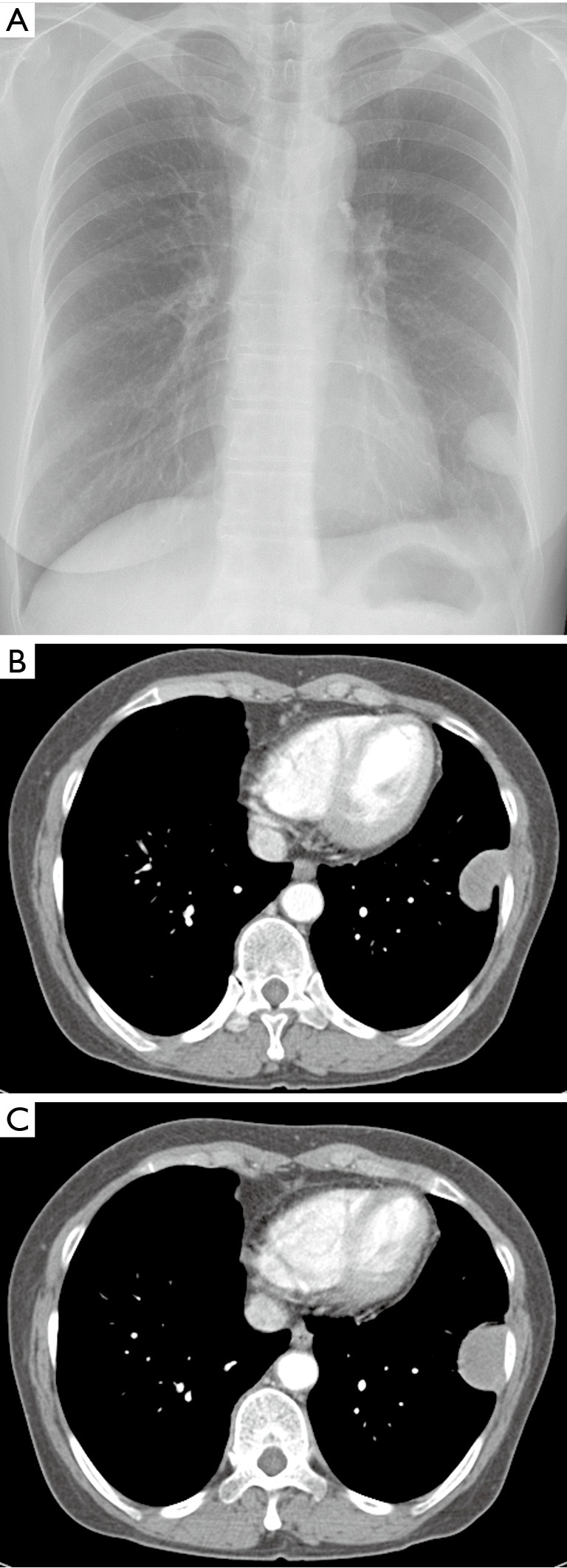
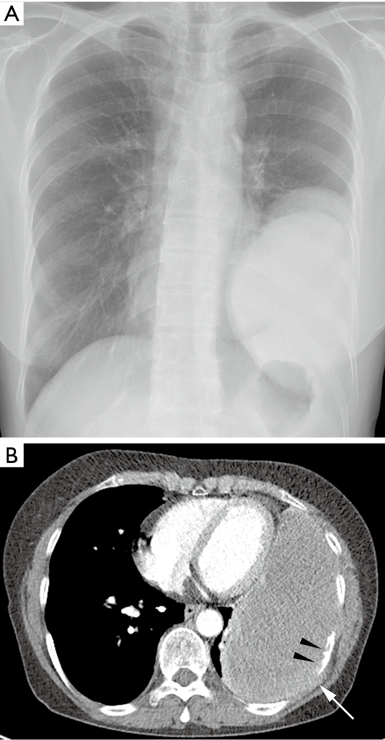
Localized malignant pleural mesothelioma (LMPM) is a rare tumor with good prognosis by surgical resection. We report an atypical case of malignant pleural sarcomatoid mesothelioma (SM) in an asymptomatic 65-year-old woman, who had no history of exposure to asbestos. She presented with a small pleural mass without pleural effusion and was misdiagnosed as a benign localized fibrous tumor (BLFT) on pathologic examination through a surgical tumor specimen. However, seven months later, the patient returned with serious cancerous symptoms. A large recurrent tumor mass was found within the chest wall invading at the old surgical resection site. SM, a subtype of LMPM, was confirmed with histopathogy and immunohistochemisty. In conclusion, malignant pleural mesothelioma (MPM) can present with typical radiologic finding similar to a BLFT, and has a wide histopathologic presentation in biopsy specimen. A thorough pathologic investigation should be attempted even when a pleural mass resembles benign, localized, and small on radiologic studies.
Keywords: Pleura; computed tomography (CT); lobectomy; mesothelioma; pathology.
Conflict of interest statement
Figures

Similar articles
-
Massive localized malignant pleural mesothelioma (LMPM): manifestations on computed tomography in 6 cases.Int J Clin Exp Med. 2015 Oct 15;8(10):18367-74. eCollection 2015. Int J Clin Exp Med. 2015. PMID: 26770440 Free PMC article.
-
A Case of Pulmonary Embolism with Sarcomatoid Malignant Pleural Mesothelioma with Long-Term Pleural Effusion.Onco Targets Ther. 2021 Jul 16;14:4231-4237. doi: 10.2147/OTT.S315869. eCollection 2021. Onco Targets Ther. 2021. PMID: 34295165 Free PMC article.
-
[Localized fibrous mesothelioma with bloody pleural effusion; report of a case].Kyobu Geka. 2004 Feb;57(2):159-62. Kyobu Geka. 2004. PMID: 14978915 Japanese.
-
Benign localized fibrous tumor of the pleura: report of 25 new cases.Thorac Cardiovasc Surg. 2012 Oct;60(7):468-73. doi: 10.1055/s-0031-1295519. Epub 2012 Jan 3. Thorac Cardiovasc Surg. 2012. PMID: 22215500 Review.
-
Solitary cerebellar metastasis of malignant pleural mesothelioma: case report.Surg Neurol. 2004 Feb;61(2):174-8; discussion 178-9. doi: 10.1016/s0090-3019(03)00448-8. Surg Neurol. 2004. PMID: 14751636 Review.
Cited by
-
Pleural mesothelioma (PMe): The evolving molecular knowledge of a rare and aggressive cancer.Mol Oncol. 2024 Apr;18(4):797-814. doi: 10.1002/1878-0261.13591. Epub 2024 Mar 8. Mol Oncol. 2024. PMID: 38459714 Free PMC article. Review.
-
Head to head comparison of 68Ga-DOTA-FAPI-04 vs 18F-FDG PET/CT in the evaluation of primary extrapulmonary tumors in the chest.Eur Radiol. 2024 Mar;34(3):1960-1970. doi: 10.1007/s00330-023-10130-3. Epub 2023 Sep 5. Eur Radiol. 2024. PMID: 37668694
-
Localized malignant pleural mesothelioma mimicking an anterior mediastinal tumor.Eur J Radiol Open. 2019 Jan 31;6:72-77. doi: 10.1016/j.ejro.2019.01.006. eCollection 2019. Eur J Radiol Open. 2019. PMID: 30740474 Free PMC article.
-
Localized malignant mesothelioma, an unusual and poorly characterized neoplasm of serosal origin: best current evidence from the literature and the International Mesothelioma Panel.Mod Pathol. 2020 Feb;33(2):281-296. doi: 10.1038/s41379-019-0352-3. Epub 2019 Sep 4. Mod Pathol. 2020. PMID: 31485011 Free PMC article. Review.
-
Malignant Pleural Mesothelioma, Biphasic Type: An Unusual and Insidious Case of Rapidly Progressive Small Blue Cell Tumor.Cureus. 2018 Jun 6;10(6):e2749. doi: 10.7759/cureus.2749. Cureus. 2018. PMID: 30109162 Free PMC article.
References
-
- American Cancer Society. Malignant Mesothelioma. 2015 Copyright American Cancer Society. Available online: www.cancer.org/malignant-mesothelioma-pdf
Publication types
LinkOut - more resources
Full Text Sources
Other Literature Sources