Case Reports
. 2016 Jun;8(6):E412-6.
doi: 10.21037/jtd.2016.04.57.

Pericardial synovial sarcoma presenting with large recurrent pericardial effusion

Affiliations
Case Reports

Pericardial synovial sarcoma presenting with large recurrent pericardial effusion

Hyo Chul Youn et al. J Thorac Dis. 2016 Jun.

Abstract

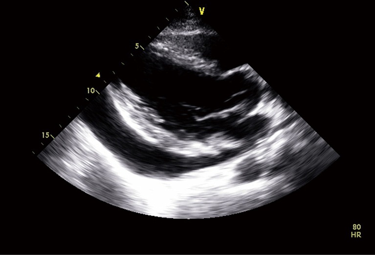
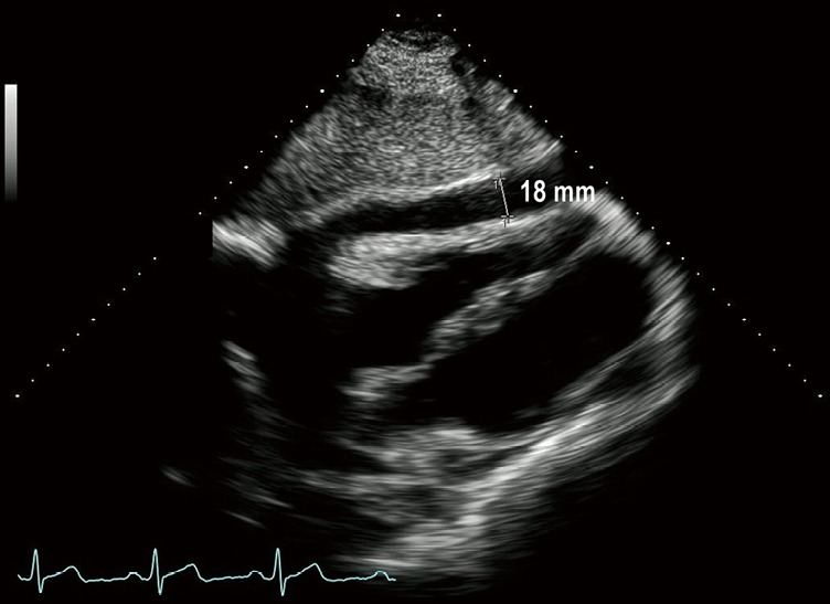
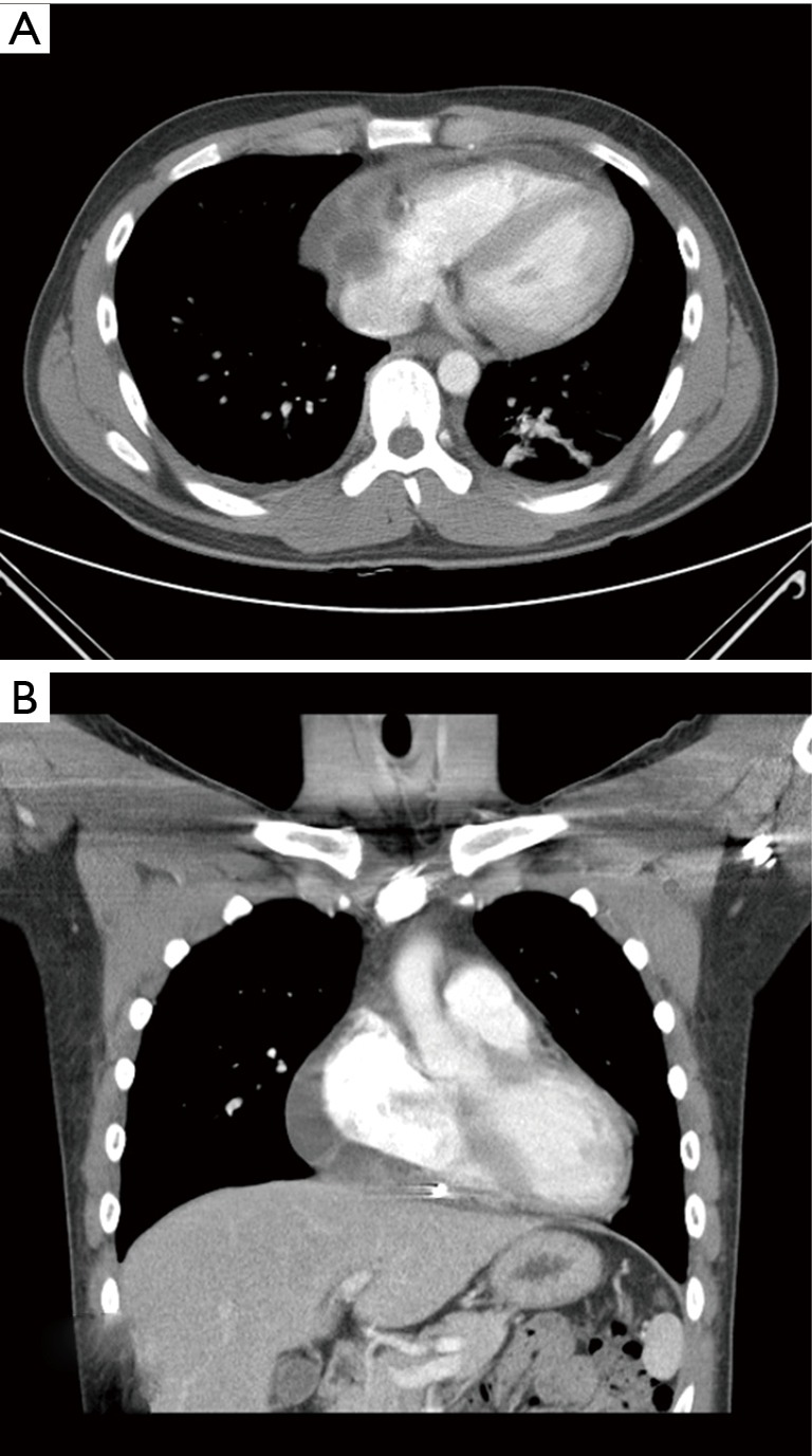
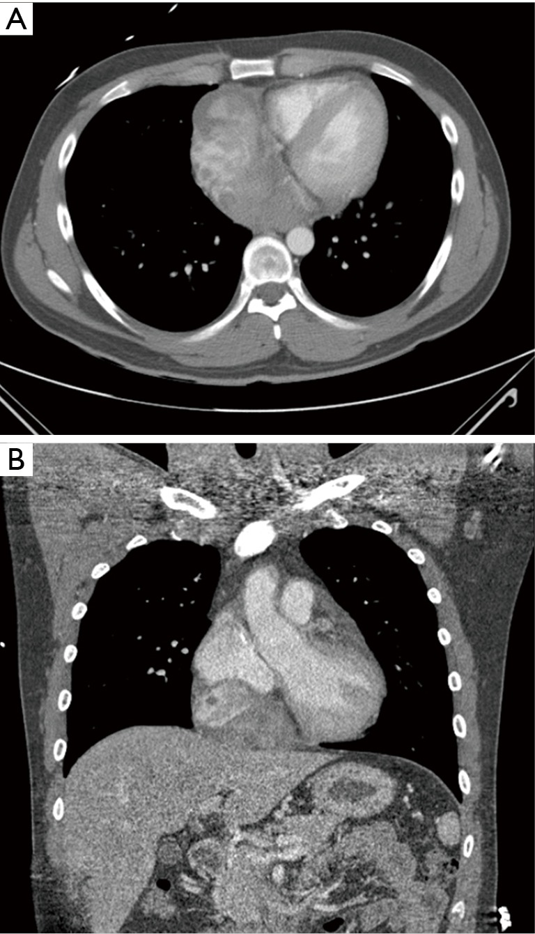
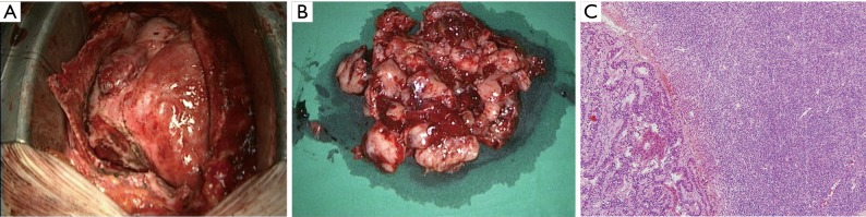
Primary pericardial synovial sarcoma is an extremely rare disease with a dismal prognosis. Its main presenting symptoms are a large pericardial effusion, signs of cardiac tamponade, and visualization of a pericardial mass on echocardiography. However, the systemic symptoms of fever, cough, and night sweats may present a clinical picture without any apparent pericardial mass on diagnostic imaging, potentially impeding the diagnosis. We report the case of a 34-year-old patient with fever and recurrent pericardial effusion for 2 years, who was diagnosed with primary pericardial synovial sarcoma after 2-year follow-up echocardiography.

Keywords: Synovial sarcoma; heart; pericardial effusion; pericardium.


Conflict of interest statement

Conflicts of Interest: The authors have no conflicts of interest to declare.

Figures

Figure 1
Figure 1
Parasternal long-axis echocardiographic view showing a large amount of pericardial effusion.
Figure 2
Figure 2
Echocardiography showing a mild amount of pericardial effusion (1.8 cm on the right side).
Figure 3
Figure 3
Chest CT findings. Axial (A) and coronal (B) contrast-enhanced CT images, showing localized pericardial effusion with septation and the thickened pericardium.
Figure 4
Figure 4
Cardiac chest CT angiographic findings. Axial (A) and coronal (B) contrast-enhanced CT images show a large ovoid mass on the right inferior side of the pericardial space, compressing the right atrium.
Figure 5
Figure 5
Surgical excision was performed. (A) The tumor is located in the pericardium; (B) gross specimen shows a large, well-demarcated tumor; (C) photomicrograph shows biphasic synovial sarcoma in which epithelial and spindle-cell components are present (Haematoxylin & eosin, ×100).

Similar articles

Cited by

References

    1. Yoshino M, Sekine Y, Koh E, et al. Pericardial synovial sarcoma: a case report and review of the literature. Surg Today 2014;44:2167-73. 10.1007/s00595-013-0720-4 - DOI - PubMed
    1. Ohzeki M, Fujita S, Miyazaki H, et al. A patient with primary pericardial synovial sarcoma who presented with cardiac tamponade: a case report and review of the literature. Intern Med 2014;53:595-601. 10.2169/internalmedicine.53.1749 - DOI - PubMed
    1. Cheng Y, Sheng W, Zhou X, et al. Pericardial synovial sarcoma, a potential for misdiagnosis: clinicopathologic and molecular cytogenetic analysis of three cases with literature review. Am J Clin Pathol 2012;137:142-9. 10.1309/AJCP34ZVFLAUTMGL - DOI - PubMed
    1. Restrepo CS, Vargas D, Ocazionez D, et al. Primary pericardial tumors. Radiographics 2013;33:1613-30. 10.1148/rg.336135512 - DOI - PubMed
    1. Burke AP, Cowan DN, Virmani R. Primary Sarcomas of the Heart. Cancer 1992;69;387-95. 10.1002/1097-0142(19920115)69:2<387::AID-CNCR2820690219>3.0.CO;2-N - DOI - PubMed

Publication types

LinkOut - more resources