. 2016 May 1;6(5):878-93.
eCollection 2016.

Prolactin-induced protein as a potential therapy response marker of adjuvant chemotherapy in breast cancer patients

Affiliations

Prolactin-induced protein as a potential therapy response marker of adjuvant chemotherapy in breast cancer patients

Karolina Jablonska et al. Am J Cancer Res. .

Abstract

Many studies are dedicated to exploring the molecular mechanisms of chemotherapy-resistance in breast cancer (BC). Some of them are focused on searching for candidate genes responsible for this process. The aim of this study was typing the candidate genes associated with the response to standard chemotherapy in the case of invasive ductal carcinoma. Frozen material from 28 biopsies obtained from IDC patients with different responses to chemotherapy were examined using gene expression microarray, Real-Time PCR (RT-PCR) and Western blot (WB). Based on the microarray results, further analysis of candidate gene expression was evaluated in 120 IDC cases by RT-PCR and in 224 IDC cases by immunohistochemistry (IHC). The results were correlated with clinical outcome and molecular subtype of the BC. Gene expression microarray revealed Prolactin-Induced Peptide (PIP) as a single gene differentially expressed in BC therapy responder or non-responder patients (p <0.05). The level of PIP expression was significantly higher in the BC therapy responder group than in the non-responder group at mRNA (p=0.0092) and protein level (p=0.0256). Expression of PIP mRNA was the highest in estrogen receptor positive (ER+) BC cases (p=0.0254) and it was the lowest in triple negative breast cancer (TNBC) (p=0.0336). Higher PIP mRNA expression was characterized by significantly longer disease free survival (DFS, p=0.0093), as well as metastasis free survival (MFS, p=0.0144). Additionally, PIP mRNA and PIP protein expression levels were significantly higher in luminal A than in other molecular subtypes and TNBC. Moreover significantly higher PIP expression was observed in G1, G2 vs. G3 cases (p=0.0027 and p=0.0013, respectively). Microarray analysis characterized PIP gene as a candidate for BC standard chemotherapy response marker. Analysis of clinical data suggests that PIP may be a good prognostic and predictive marker in IDC patients. Higher levels of PIP were related to longer DFS and MFS but not with OS.

Keywords: PIP; adjuvant chemotherapy; breast cancer.


Figures
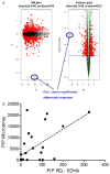
Figure 1
Figure 1
A. The analysis of genes from the expression arrays. The MA and Volcano plots showed the spread of the differentially expressed genes and PIP gene as the only gene differentially expressed in patients with non-responder or responder status. B. Real-time PCR analysis of PIP gene expression significantly correlated positively with microarray method (r=0.66; p=0.001).
Figure 2
Figure 2
A. PIP mRNA relative expression (RQ) is significantly higher in the breast cancer (BC) therapy responder group than in the non-responder group (**p=0.0092). B. Densitometry analysis of IDC cases of therapy responder group (R) as compared to non-responder (NR) demonstrates significantly higher PIP protein level in the group of R vs. NR (*p=0.0256). C. Western blot of 4 responders and 6 non-responders BC samples shows the differences in the amount of PIP protein.
Figure 3
Figure 3
A. BC patients with higher PIP expression are characterized by a significantly longer disease free survival (DFS; **p=0.0093), B. BC patients with higher PIP expression are characterized by longer metastasis free survival (MFS; *p=0.0144).
Figure 4
Figure 4
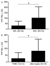
A. PIP mRNA relative expression (RQ) is significantly higher in the BC cases with positive estrogen receptor status ER+ than in ER- cases (*p=0.0254), B. PIP mRNA relative expression (RQ) is significantly lower in the TNBC (*p=0.0336).
Figure 5
Figure 5
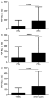
Association of PIP mRNA level with clinicopathological features. A. PIP mRNA relative expression (RQ) is significantly higher in the group of ER+ than in ER- IDC cases (****p <0.0001). B. PR+ cases showed significantly higher levels of PIP mRNA expression than PR- IDC (****p <0.0001). C. TNBC cases were characterized by significantly lower PIP mRNA RQ than in other cases (****p <0.0001).
Figure 6
Figure 6
Association of PIP mRNA expression with BC molecular subtypes. PIP mRNA RQ was significantly higher in luminal A than in other molecular subtypes and to the TNBC as well as in other molecular subtypes to the TNBC (*p=0.0268, ***p=0.0005 and **p=0.0041 respectively).
Figure 7
Figure 7
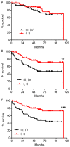
Univariate survival analysis of PIP mRNA of 120 IDC patients. (A) Mantel-Cox test demonstrates no significant association of overall survival with pTNM staging. (B) Lower pTNM staging correlates significantly with longer disease free survival (DFS; III-IV vs. I-II; **p=0.0046) as well as with (C) longer metastasis free survival (MFS; III-IV vs. I-II; ***p=0.0007).
Figure 8
Figure 8
Survival analysis of PIP mRNA. (A) PIP mRNA expression showed no significant association with overall survival. (B) Higher level of PIP mRNA expression strongly correlates with longer disease free survival (DFS; ****p <0.0001) as well as with (C) longer metastasis free survival (MFS; ***p=0.0006).
Figure 9
Figure 9
Survival analysis with using online analysis tool on 1,115 cases of BC. The good prognostic effect of high PIP expression is related to longer OS [31].
Figure 10
Figure 10
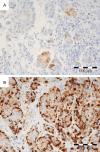
Immunohistochemical expression pattern of PIP in IDC cells. Expression of PIP was localized only in cytoplasm of tumor cells. A. Low PIP expression. B. High intensity of PIP expression. Magnification ×200.
Figure 11
Figure 11
PIP expression in BC cells. Significantly higher PIP expression level is observed in G1 and G2 as compared to G3 cases (* p=0.027 and **p=0.0013, respectively).
Figure 12
Figure 12
Association of PIP expression (IHC) with clinicopathological traits. A. PR positive (PR+) tumors show significantly higher level of PIP expression than PR negative (PR-) cases (*p <0.05). B. Triple negative (TNBC) cases exhibited lower level of PIP expression as compared with other molecular subtypes (***p <0.001). C. PIP expression negatively correlates with Ki-67 proliferation marker in BC samples (r=-0.254; p=0.0001).
Figure 13
Figure 13
Association of PIP (IHC) expression with BC molecular subtypes. Significantly higher PIP expression in luminal A as compared with the other molecular subtypes and with the TNBC as well as other molecular subtypes to TNBC (***p=0.0003; ****p <0.0001; *p=0.0343, respectively).
Figure 14
Figure 14
The univariate analyses of 224 cases of IDC patients. (A) Lower pTNM staging (III-IV vs. I-II **p=0.0075) demonstrated significant association with the longer overall survival, (B) Lower malignancy grade (G1 and G2 vs. G3; **p=0.0037) correlates significantly with the longer OS and (C) low Ki-67 expression in the BC cells correlates significantly with the longer OS (≤25%; *p=0.0469). (D) PIP expression level (IHC) in BC cells showed no significant association with OS. The IHC cut-off scores for OS: 0-3 pts vs. 4-12 pts.

References

    1. Siegel R, Naishadham D, Jemal A. Cancer statistics, 2013. CA Cancer J Clin. 2013;63:11–30. - PubMed
    1. Goldhirsch A, Wood WC, Coates AS, Gelber RD, Thürlimann B, Senn HJ, members P. Strategies for subtypes--dealing with the diversity of breast cancer: highlights of the St. Gallen International Expert Consensus on the Primary Therapy of Early Breast Cancer 2011. Ann Oncol. 2011;22:1736–1747. - PMC - PubMed
    1. Goldhirsch A, Ingle JN, Gelber RD, Coates AS, Thürlimann B, Senn HJ, members P. Thresholds for therapies: highlights of the St Gallen International Expert Consensus on the primary therapy of early breast cancer 2009. Ann Oncol. 2009;20:1319–1329. - PMC - PubMed
    1. Mazoujian G, Pinkus GS, Davis S, Haagensen DE. Immunohistochemistry of a gross cystic disease fluid protein (GCDFP-15) of the breast. A marker of apocrine epithelium and breast carcinomas with apocrine features. Am J Pathol. 1983;110:105–112. - PMC - PubMed
    1. Pagani A, Sapino A, Eusebi V, Bergnolo P, Bussolati G. PIP/GCDFP-15 gene expression and apocrine differentiation in carcinomas of the breast. Virchows Arch. 1994;425:459–465. - PubMed

LinkOut - more resources