

Retinal Macroglial Responses in Health and Disease
- PMID: 27294114
- PMCID: PMC4887628
- DOI: 10.1155/2016/2954721
Retinal Macroglial Responses in Health and Disease
Abstract
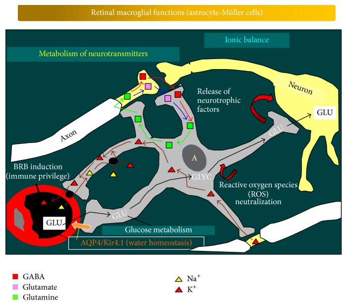
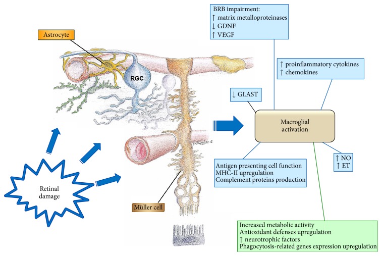
Due to their permanent and close proximity to neurons, glial cells perform essential tasks for the normal physiology of the retina. Astrocytes and Müller cells (retinal macroglia) provide physical support to neurons and supplement them with several metabolites and growth factors. Macroglia are involved in maintaining the homeostasis of extracellular ions and neurotransmitters, are essential for information processing in neural circuits, participate in retinal glucose metabolism and in removing metabolic waste products, regulate local blood flow, induce the blood-retinal barrier (BRB), play fundamental roles in local immune response, and protect neurons from oxidative damage. In response to polyetiological insults, glia cells react with a process called reactive gliosis, seeking to maintain retinal homeostasis. When malfunctioning, macroglial cells can become primary pathogenic elements. A reactive gliosis has been described in different retinal pathologies, including age-related macular degeneration (AMD), diabetes, glaucoma, retinal detachment, or retinitis pigmentosa. A better understanding of the dual, neuroprotective, or cytotoxic effect of macroglial involvement in retinal pathologies would help in treating the physiopathology of these diseases. The extensive participation of the macroglia in retinal diseases points to these cells as innovative targets for new drug therapies.
Figures
Similar articles
-
Purinergic signaling in retinal degeneration and regeneration.Neuropharmacology. 2016 May;104:194-211. doi: 10.1016/j.neuropharm.2015.05.005. Epub 2015 May 19. Neuropharmacology. 2016. PMID: 25998275 Review.
-
670nm light treatment following retinal injury modulates Müller cell gliosis: Evidence from in vivo and in vitro stress models.Exp Eye Res. 2018 Apr;169:1-12. doi: 10.1016/j.exer.2018.01.011. Epub 2018 Feb 3. Exp Eye Res. 2018. PMID: 29355737
-
Endothelin-2 Injures the Blood-Retinal Barrier and Macroglial Müller Cells: Interactions with Angiotensin II, Aldosterone, and NADPH Oxidase.Am J Pathol. 2018 Mar;188(3):805-817. doi: 10.1016/j.ajpath.2017.11.009. Epub 2017 Dec 15. Am J Pathol. 2018. PMID: 29248456
-
Ebselen by modulating oxidative stress improves hypoxia-induced macroglial Müller cell and vascular injury in the retina.Exp Eye Res. 2015 Jul;136:1-8. doi: 10.1016/j.exer.2015.04.015. Epub 2015 Apr 24. Exp Eye Res. 2015. PMID: 25912997
-
Glia-neuron interactions in the mammalian retina.Prog Retin Eye Res. 2016 Mar;51:1-40. doi: 10.1016/j.preteyeres.2015.06.003. Epub 2015 Jun 23. Prog Retin Eye Res. 2016. PMID: 26113209 Review.
Cited by
-
Contralateral Astrocyte Response to Acute Optic Nerve Damage Is Mitigated by PANX1 Channel Activity.Int J Mol Sci. 2023 Oct 27;24(21):15641. doi: 10.3390/ijms242115641. Int J Mol Sci. 2023. PMID: 37958624 Free PMC article.
-
Intravitreal Injection of PACAP Attenuates Acute Ocular Hypertension-Induced Retinal Injury Via Anti-Apoptosis and Anti-Inflammation in Mice.Invest Ophthalmol Vis Sci. 2022 Mar 2;63(3):18. doi: 10.1167/iovs.63.3.18. Invest Ophthalmol Vis Sci. 2022. PMID: 35293951 Free PMC article.
-
Alterations of Ocular Hemodynamics Impair Ophthalmic Vascular and Neuroretinal Function.Am J Pathol. 2018 Mar;188(3):818-827. doi: 10.1016/j.ajpath.2017.11.015. Epub 2018 Jan 5. Am J Pathol. 2018. PMID: 29309745 Free PMC article.
-
O-Linked β-N-acetylglucosamine (O-GlcNAc) modification: a new pathway to decode pathogenesis of diabetic retinopathy.Clin Sci (Lond). 2018 Jan 19;132(2):185-198. doi: 10.1042/CS20171454. Print 2018 Jan 31. Clin Sci (Lond). 2018. PMID: 29352075 Free PMC article. Review.
-
Peripapillary Intravitreal Injection Improves AAV-Mediated Retinal Transduction.Mol Ther Methods Clin Dev. 2020 Mar 30;17:647-656. doi: 10.1016/j.omtm.2020.03.018. eCollection 2020 Jun 12. Mol Ther Methods Clin Dev. 2020. PMID: 32300611 Free PMC article.
References
-
- Kandel E. R., Schwartz J. H., Jessell T. M. Neuronas y conducta. In: Kandel E. R., Schwartz J. H., Jessell T. M., editors. Principios de Neurociencia. Madrid, Spain: McGraw-Hill/Interamericana; 2001. pp. 19–35.
Publication types
MeSH terms
Substances
LinkOut - more resources
Full Text Sources
Other Literature Sources
