Review
. 2016 Sep;116(9):1595-625.
doi: 10.1007/s00421-016-3411-1. Epub 2016 Jun 13.

Genetic variation and exercise-induced muscle damage: implications for athletic performance, injury and ageing

Affiliations
Review

Genetic variation and exercise-induced muscle damage: implications for athletic performance, injury and ageing

Philipp Baumert et al. Eur J Appl Physiol. 2016 Sep.

Abstract

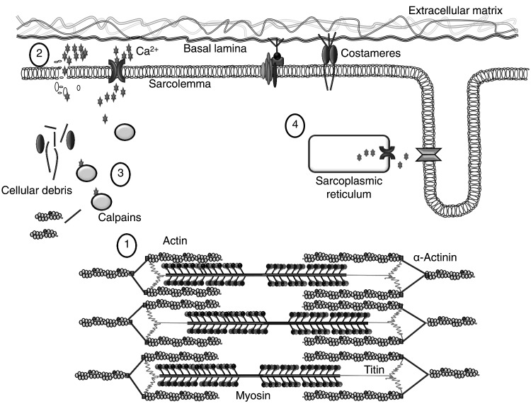
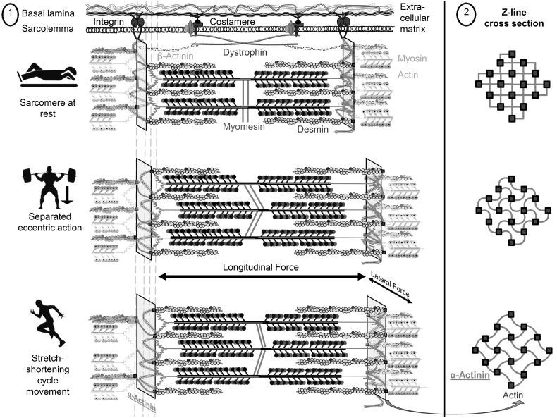
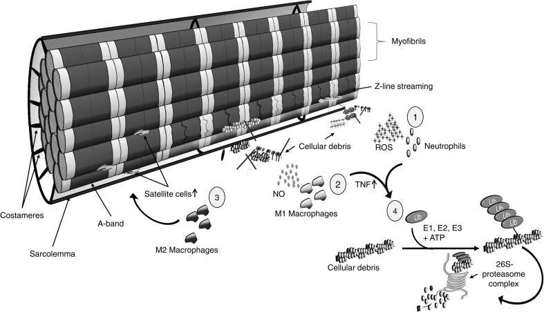
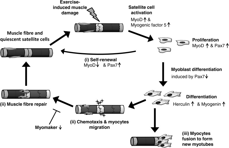
Prolonged unaccustomed exercise involving muscle lengthening (eccentric) actions can result in ultrastructural muscle disruption, impaired excitation-contraction coupling, inflammation and muscle protein degradation. This process is associated with delayed onset muscle soreness and is referred to as exercise-induced muscle damage. Although a certain amount of muscle damage may be necessary for adaptation to occur, excessive damage or inadequate recovery from exercise-induced muscle damage can increase injury risk, particularly in older individuals, who experience more damage and require longer to recover from muscle damaging exercise than younger adults. Furthermore, it is apparent that inter-individual variation exists in the response to exercise-induced muscle damage, and there is evidence that genetic variability may play a key role. Although this area of research is in its infancy, certain gene variations, or polymorphisms have been associated with exercise-induced muscle damage (i.e. individuals with certain genotypes experience greater muscle damage, and require longer recovery, following strenuous exercise). These polymorphisms include ACTN3 (R577X, rs1815739), TNF (-308 G>A, rs1800629), IL6 (-174 G>C, rs1800795), and IGF2 (ApaI, 17200 G>A, rs680). Knowing how someone is likely to respond to a particular type of exercise could help coaches/practitioners individualise the exercise training of their athletes/patients, thus maximising recovery and adaptation, while reducing overload-associated injury risk. The purpose of this review is to provide a critical analysis of the literature concerning gene polymorphisms associated with exercise-induced muscle damage, both in young and older individuals, and to highlight the potential mechanisms underpinning these associations, thus providing a better understanding of exercise-induced muscle damage.

Keywords: Creatine kinase; Delayed onset muscle soreness; Elderly; Exercise-induced muscle damage; Single nucleotide polymorphism.


Figures

Fig. 1
Fig. 1
Initial phase of exercise-induced muscle damage. Due to different abilities of each sarcomere to resist eccentric actions, some of the sarcomeres will be stretched beyond the optimum overlap of actin and myosin filaments, resulting in Z-line streaming (Morgan, 1990) (1). This is accompanied by increased permeability of the sarcolemma (2). Extracellular Ca2+ influx into the muscle fibre activates different Ca2+-sensitive proteases (calpains). Calpain activation leads to proteolysis of cytoskeletal and costameric proteins (Thiebaud 2012) (3). However, a failure of excitation–contraction coupling also seems to play an important role in strength loss following strenuous exercise, as murine muscle exposed to caffeine revealed an attenuated loss of muscle strength (Warren et al. 1993) (4). Figure adapted from Hyldahl and Hubal (2014)
Fig. 2
Fig. 2
Proposed changes in sarcomere structure during stretch–shortening cycle movements and purely eccentric actions, focussing on α-actinin (highlighted in red and underlined). The left-hand side shows the sarcomere longitudinally in a quasi-3D model at rest, and the α-actinin elongation during purely eccentric actions, and stretch–shortening cycle movements (1). The right-hand side illustrates the sarcomere cross-section at the level of the Z-line (2). At rest, α-actinin is set to roughly 90° between the antiparallel actin filaments, while under active tension, the space between the antiparallel actin filaments increases and α-actinin is stretched to a basket-weave lattice (Gautel 2011). Alpha-actinin is thought to play a key role in the longitudinal (via the anchoring of actin filaments to the Z-line) and lateral (via costamere fibre-to-fibre interaction) transmission of muscle fibre force (Hughes et al. ; Yang and Xu 2012). Moreover, human type II muscle fibres from ACTN3 XX homozygotes (where α-actinin-3 deficiency is compensated by the presence of α-actinin-2) are less stiff than type II muscle fibres from ACTN3 R-allele carriers (Broos et al. 2012). Thus, it is likely that α-actinin-2 is able to store more energy than α-actinin-3 during the active stretch phase of the stretch–shortening cycle, which is released during the shortening phase (Kjær ; Yang and Xu 2012). We propose that stretch–shortening cycle movements increase the actin filament spacing to a greater extent compared to purely eccentric actions, thus elongating α-actinin to become almost completely straight at peak eccentric force. Individuals with α-actinin-3 deficiency (ACTN3 XX homozygotes) might, therefore, benefit from having a more elastic Z-line during stretch–shortening cycle movements compared to R-allele carriers (Broos et al. 2012), resulting in a reduced damage response to stretch–shortening movements (Venckunas et al. 2012). Figure adapted from Gautel (2011) (color figure online)
Fig. 3
Fig. 3
The secondary phase of muscle damage. Leucocytes infiltrate the site of myotrauma (Tidball 2005). Firstly, neutrophils migrate to damaged muscle fibres and produce reactive oxygen species (ROS) to degrade cellular debris (Suzuki et al. 1996) (1). Neutrophils are substituted by macrophages within 24 h (Malm et al. 2000), with M1 macrophages removing cellular debris by producing cytotoxic levels of nitric oxide (NO) (2). In the latter stage of muscle damage, a shift from M1 to M2 macrophages is associated with the activation of satellite cells and the subsequent regeneration of muscle fibres (Tidball 2011) (3). Neutrophils and macrophages also express tumour necrosis factor (TNF), which activates the ubiquitin–proteasome pathway (Tidball and Villalta 2010) (4). This pathway regulates proteolysis by attaching ubiquitin polymers (Ub) to cellular debris via three different types of enzymes (E1–E3 ligases). As a result, these ubiquitin-marked proteins will be degraded by the 26S-proteasome complex (Reid 2005)
Fig. 4
Fig. 4
The cycle of skeletal muscle fibre regeneration following exercise-induced muscle damage. This cycle is mediated by satellite cells, which are activated following stressful physiological conditions such as exercise-induced muscle damage (Grobler et al. 2004). Activated satellite cells initially up-regulate two different myogenic regulatory factors, MyoD and myogenic factor-5 and, during the proliferation, paired box protein 7 (Pax7). If satellite cells return to quiescence and restore the population of satellite cells, MyoD will be down-regulated (i). However, subsequent cell differentiation is accompanied with down-regulation of Pax7/3. During this early differentiation stage, herculin and myogenin are up-regulated. Myoblasts differentiate into myocytes and then eventually migrate to the site of injury and support the repair process by increasing the nuclei-to-cytoplasm ratio (ii). Different chemotactic gradients, including a large number of chemokines, support the migration to the region of injury. A recent investigation in mice revealed that the absence of myomaker, which is expressed on the cell surface of myoblasts, leads to inhibition of myoblast fusion (Millay et al. 2013). Alternatively, the myocytes fuse with other myogenic cells to form myotubes, thus generating new fibres to replace damaged myofibres (iii). Figure adapted from Tidball (2011) and Al-Shanti and Stewart (2009)

Similar articles

Cited by

References

    1. Ahmetov II, Naumov VA, Donnikov AE, Maciejewska-Karłowska A, Kostryukova ES, Larin AK, Maykova EV, Alexeev DG, Fedotovskaya ON, Generozov EV. SOD2 gene polymorphism and muscle damage markers in elite athletes. Free Radical Res. 2014;48(8):948–955. - PubMed
    1. Akimoto AK, Miranda-Vilela AL, Alves PC, Pereira LC, Lordelo GS, Hiragi Cde O, da Silva IC, Grisolia CK, Klautau-Guimaraes Mde N. Evaluation of gene polymorphisms in exercise-induced oxidative stress and damage. Free Radical Res. 2010;44(3):322–331. - PubMed
    1. Allen NE, Davey GK, Key TJ, Zhang S, Narod SA. Serum insulin-like growth factor I (IGF-I) concentration in men is not associated with the cytosine-adenosine repeat polymorphism of the IGF-I gene. Cancer Epidem Biomark. 2002;11(3):319–320. - PubMed
    1. Allen DG, Whitehead NP, Yeung EW. Mechanisms of stretch-induced muscle damage in normal and dystrophic muscle: role of ionic changes. J Physiol. 2005;567(3):723–735. - PMC - PubMed
    1. Al-Shanti N, Stewart CE. Ca2+/calmodulin-dependent transcriptional pathways: potential mediators of skeletal muscle growth and development. Biol Rev. 2009;84(4):637–652. - PubMed

MeSH terms