. 2016 Jun 13;11(6):e0157453.
doi: 10.1371/journal.pone.0157453. eCollection 2016.

TASK-1 Regulates Apoptosis and Proliferation in a Subset of Non-Small Cell Lung Cancers

Affiliations

TASK-1 Regulates Apoptosis and Proliferation in a Subset of Non-Small Cell Lung Cancers

Katharina Leithner et al. PLoS One. .

Abstract

Lung cancer is the leading cause of cancer deaths worldwide; survival times are poor despite therapy. The role of the two-pore domain K+ (K2P) channel TASK-1 (KCNK3) in lung cancer is at present unknown. We found that TASK-1 is expressed in non-small cell lung cancer (NSCLC) cell lines at variable levels. In a highly TASK-1 expressing NSCLC cell line, A549, a characteristic pH- and hypoxia-sensitive non-inactivating K+ current was measured, indicating the presence of functional TASK-1 channels. Inhibition of TASK-1 led to significant depolarization in these cells. Knockdown of TASK-1 by siRNA significantly enhanced apoptosis and reduced proliferation in A549 cells, but not in weakly TASK-1 expressing NCI-H358 cells. Na+-coupled nutrient transport across the cell membrane is functionally coupled to the efflux of K+ via K+ channels, thus TASK-1 may potentially influence Na+-coupled nutrient transport. In contrast to TASK-1, which was not differentially expressed in lung cancer vs. normal lung tissue, we found the Na+-coupled nutrient transporters, SLC5A3, SLC5A6, and SLC38A1, transporters for myo-inositol, biotin and glutamine, respectively, to be significantly overexpressed in lung adenocarcinomas. In summary, we show for the first time that the TASK-1 channel regulates apoptosis and proliferation in a subset of NSCLC.


Conflict of interest statement

Competing Interests: Our work was funded by the Oesterreichische Nationalbank (Anniversary Fund, project number 12713). The Oesterreichische Nationalbank does not have any commercial interests associated with the funded projects including our project. The Oesterreichische Nationalbank is owned by the federal government of Austria, and the Anniversary Fund supports only independent research from very diverse research fields. The projects are reviewed by independent, international researchers (peer-review) and the results obtained are not in any form utilized by the Oesterreichische Nationalbank, nor does the Oesterreichische Nationalbank have any impact on the aims of the projects. This does not alter our adherence to PLOS ONE policies on sharing data and materials.

Figures

Fig 1
Fig 1. Expression of TASK-1 in lung cancer cell lines.
(A) TASK-1 expression in eight different NSCLC cell lines. A representative immunoblot and mean densitometry values are shown. A549 cells, present on all immunoblots, served as a reference for normalization. Bottom: TASK-1 mRNA levels were assessed by quantitative RT-PCR. Beta-actin (ACTB) was used as a reference gene. (B) TASK-3 protein and mRNA levels in eight different NSCLC cell lines. (A,B) Results are mean +/- SEM from three to four independent samples. Group comparisons were calculated with one-group Student′s t-test. (C,D) A549 cells were transfected with non-silencing siRNA (c), TASK-1 siRNA (T) or left untreated (u). (C) Representative immunoblot using a TASK-1 antibody in the presence or absence of a specific blocking peptide is shown. TASK-1 appears at a molecular weight of 52 kDa, the additional faint band at 46 kDa might represent the unglycolsylated form. (D) TASK-1 mRNA levels 48 hours after transfection with TASK-1 siRNA assessed by quantitative RT-PCR. (E) Representative immunoblots of TASK-1 in A549 cells and H358 cells at different time intervals after transfection. Data are shown as mean +/- SEM from n = 3 independent experiments. * P<0.05, ** P<0.01, *** P<0.001.
Fig 2
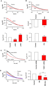
Fig 2. TASK-1 is functional and contributes to setting the resting membrane potential in A549 cells.
(A) Representative recordings of a non-inactivating K+ current (IKN) in A549 cells using the whole cell patch clamp technique under acidic and basic conditions, and graphs summarizing the mean current at 0 mV. I/I0 is the current in the presence of low (6.3) or high (8.3) pH expressed as a fraction of the current before treatment. The non-inactivating current is reduced under acidic and increased under basic conditions. (B) Representative recordings and a graph summarizing the non-inactivating K+ current (IKN) and the resting membrane potential (Em) in the presence and absence of the TASK-1 inhibitor anandamide (Ana, 10 μM) at pH 7.3 in A549 cells. I/I0 is the current in the presence of anandamide expressed as a fraction of the current before anandamide treatment. Anandamide reduced the non-inactivating K+ current and led to depolarization of the cell membrane towards more positive values. (C) Representative recordings of the non-inactivating K+ current (IKN) and bar graph summarizing the mean current at 0 mV in A549 cells transfected with TASK-1 siRNA or control siRNA at pH 7.3. (D) Representative recordings of the non-inactivating K+ current (IKN) and the normalized mean current at 0 mV in A549 cells under treatment with the TASK-3 blocker ruthenium red (RR, 10 μM) or ruthenium red together with anandamide (10 μM) at pH 7.3. Data are mean +/- SEM. ** P<0.01, *** P<0.001 compared with control. Ana, anandamide; RR, ruthenium red.
Fig 3
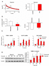
Fig 3. Hypoxia reduces the TASK-1 current in A549 cells.
(A) Left, representative recordings of the non-inactivating K+ current (IKN) in A549 cells cultured under hypoxia (1% oxygen) for three days. The detectable current lacks sensitivity to the TASK-1 inhibitor anandamide (Ana, 10 μM). Right, graph summarizing the mean current at 0 mV. (B) Non-inactivating K+ current density (IKN Density; left) and the resting membrane potential (Em; right) in A549 cells cultured under hypoxia or normoxia. (C) relative abundance of TASK-1 mRNA in A549 cells cultured under hypoxia (1% oxygen) for different time intervals measured by quantitative PCR. GLUT-1 and hexokinase 2 (HK2) were assessed as hypoxia markers. (D) Immunoblot and relative abundance of TASK-1 protein in A549 cells cultured under hypoxia (1% oxygen) measured for different time intervals. Data are mean +/- SEM. * P<0.05, *** P<0.001, n.s., not significant. Ana, anandamide; HK2, hexokinase 2; GLUT1, solute carrier family 2 (facilitated glucose transporter), member 1.
Fig 4
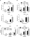
Fig 4. TASK-1 knockdown enhances apoptosis in A549 cells.
(A,B) Cells were transfected with TASK-1 siRNA or non-silencing siRNA (control siRNA). 48 hours after transfection, cells were replated, after additional 24 hours cells were treated with different stimuli for 72 hours and apoptosis was assessed by detecting cells with caspase 3 activity by FACS analysis. For apoptosis induction, cells were treated with cisplatin at different concentrations or with DMEM medium containing dialyzed serum and 10 mM or 0 mM D-glucose (balanced with metabolically inert L-glucose). (C) A549 cells were transfected and treated with different concentrations of cisplatin in the same manner as in panel A. Apoptosis was assessed by staining floating and adherent cells with Hoechst dye. Rates of nuclear fragmentation were determined in a blinded manner. (D) A549 cells were transfected in the same manner as in panel A and treated with different concentrations of cisplatin without previous replating. (A,B,D) Apoptosis was assessed by detecting cells with caspase 3 activity by FACS analysis. Results are mean +/- SEM from n = 3 independent experiments. Group comparisons were performed with Two-way ANOVA followed by Bonferroni post-hoc analysis. * P<0.05, ** P<0.01; *** P<0.001. n.s., not significant.
Fig 5
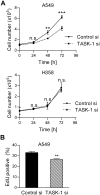
Fig 5. TASK-1 knockdown reduces proliferation in A549 cells.
(A) Cells were transfected with TASK-1 siRNA or non-silencing siRNA (Control siRNA). 48 hours after transfection cells were replated in medium with reduced serum content (1%), in order to avoid overstimulation of cells. Total cell numbers were counted each consecutive day in duplicates with electronic pulse area analysis (CASY®). Results are mean +/- SEM from n = 3 independent experiments. Group comparisons were performed with Two-way ANOVA followed by Bonferroni post-hoc analysis. (B) Cells were transfected as described and grown in serum-reduced medium (1%) for 48 hours. Mitosis was assessed using an EdU uptake assay. Results are mean +/- SEM from n = 4 independent experiments. ** P<0.01, *** P<0.001, n.s., not significant.
Fig 6
Fig 6. Expression of TASK-1 and of putative downstream effectors of TASK-1, Na+-coupled transporters in human NSCLC.
(A) TASK-1 protein was assessed in NSCLC tissue and corresponding non-involved lung from twelve patients using Western blot. Right: Densitometry values for TASK-1 in NSCLC and lungs were normalized to β-actin. (B,C) mRNA levels of members of the SLC5 family of Na+-coupled transporters and of the Na+-driven glutamine transporter SLC38A1 were assessed in a publically available GEO dataset (GDS3257) [11] published at Gene Expression Omnibus (GEO; http://www.ncbi.nlm.nih.gov/geo/) in lung adenocarcinoma samples (n = 58) and normal lungs (n = 49). The RMA (Robust Multichip Average) expression measure for mRNA abundance is in the log scale. *** P<0.001, * P<0.05.

Similar articles

Cited by

References

    1. Ramalingam SS, Owonikoko TK, Khuri FR. Lung cancer: New biological insights and recent therapeutic advances. CA Cancer J Clin. 2011;61(2):91–112. 10.3322/caac.20102 - DOI - PubMed
    1. Manegold C, Thatcher N. Survival improvement in thoracic cancer: progress from the last decade and beyond. Lung Cancer. 2007;57 Suppl 2:S3–S5. - PubMed
    1. Pardo LA, Stuhmer W. The roles of K(+) channels in cancer. Nat Rev Cancer. 2014;14(1):39–48. 10.1038/nrc3635 - DOI - PubMed
    1. Prevarskaya N, Skryma R, Shuba Y. Ion channels and the hallmarks of cancer. Trends Mol Med. 2010;16(3):107–121. 10.1016/j.molmed.2010.01.005 - DOI - PubMed
    1. Olschewski A. Targeting TASK-1 channels as a therapeutic approach. Adv Exp Med Biol. 2010;661:459–473. 10.1007/978-1-60761-500-2_30 - DOI - PubMed

MeSH terms

Substances