. 2016 Sep:101:258-71.
doi: 10.1016/j.biomaterials.2016.06.006. Epub 2016 Jun 3.

Affinity-controlled protein encapsulation into sub-30 nm telodendrimer nanocarriers by multivalent and synergistic interactions

Affiliations

Affinity-controlled protein encapsulation into sub-30 nm telodendrimer nanocarriers by multivalent and synergistic interactions

Xu Wang et al. Biomaterials. 2016 Sep.

Abstract

Novel nanocarriers are highly demanded for the delivery of heterogeneous protein therapeutics for disease treatments. Conventional nanoparticles for protein delivery are mostly based on the diffusion-limiting mechanisms, e.g., physical trapping and entanglement. We develop herein a novel linear-dendritic copolymer (named telodendrimer) nanocarrier for efficient protein delivery by affinitive coating. This affinity-controlled encapsulation strategy provides nanoformulations with a small particle size (<30 nm), superior loading capacity (>50% w/w) and maintained protein bioactivity. We integrate multivalent electrostatic and hydrophobic functionalities synergistically into the well-defined telodendrimer scaffold to fine-tune protein binding affinity and delivery properties. The ion strength and density of the charged groups as well as the structure of the hydrophobic segments are important and their combinations in telodendrimers are crucial for efficient protein encapsulation. We have conducted a series of studies to understand the mechanism and kinetic process of the protein loading and release, utilizing electrophoresis, isothermal titration calorimetry, Förster resonance energy transfer spectroscopy, bio-layer interferometry and computational methods. The optimized nanocarriers are able to deliver cell-impermeable therapeutic protein intracellularly to kill cancer cells efficiently. In vivo imaging studies revealed cargo proteins preferentially accumulate in subcutaneous tumors and retention of peptide therapeutics is improved in an orthotopic brain tumor, these properties are evidence of the improved pharmacokinetics and biodistributions of protein therapeutics delivered by telodendrimer nanoparticles. This study presents a bottom-up strategy to rationally design and fabricate versatile nanocarriers for encapsulation and delivery of proteins for numerous applications.

Keywords: Affinity-controlled encapsulation; Multivalent interactions; Nanoparticles; Protein delivery; Synergistic effects; Telodendrimers.


Figures

Fig. 1
Fig. 1
Schematic illustration of telodendrimer structures and protein encapsulation.
Fig. 2
Fig. 2
(a,b) Particle size (a) and zeta potential (b) of telodendrimers before and after BSA loading. The error is for standard deviation (n = 3). (c–h) TEM images and particle size analysis for PEG5k(ArgArg-L-C17)4 (c,f), PEG5k(ArgArg-L-CHO)4 (d,g), and PEG5k(ArgArg-L-VE)4 (e,h) NPs before (c–e) and after (f–h) BSA loading (P/T = 1/3, w/w). (i) Proposed process for in situ protein encapsulation. (j) Loading ability of telodendrimer NPs for FITC-BSA determined by an agarose gel retention assay. The feed mass ratio is 2/1 (P/T). (k) Calorimetric titration of PEG5k(ArgArg-L-VE)4 with BSA at 37 °C in PBS. (l,m) Fluorescence emission spectra (λex 439 nm) of RB-BSA, FITC-PEG5k(ArgArg-L-C17)4 and their mixture (l) and the mixture of RB-BSA and FITC-BSA without/with PEG5k(ArgArg-L-C17)4 (m).
Fig. 3
Fig. 3
(a) Schematic illustration of the association in telodendrimer solution (left) and dissociation in BSA solution (right) for the streptavidin-coated biosensors prewetted with BSA solution. (b) Kinetics for association in PEG5k(ArgArg-L-C17)4 solution (500 nM) and dissociation in PBS and BSA solutions of different concentrations. (c–e) Kinetics for association in PEG5k(ArgArg-L-C17)4 (c), PEG5k(ArgArg-L-CHO)4 (d), and PEG5k(ArgArg-L-VE)4 (e) solutions (75–600 nM) and dissociation in BSA solutions (40 mg/mL). (f) Summary of BLI results.
Fig. 4
Fig. 4
(a) Average docking energy of different subunits with BSA (average of 100 docking runs). (b) Loading efficiency of telodendrimers for FITC-BSA determined by an agarose gel retention assay. The feed mass ratio is 1/3 (P/T). (c–e) Kinetics for association in telodendrimer solutions and dissociation in BSA solution (40 mg/mL). (f–h) Fluorescence emission spectra of mixture of RB-BSA and FITC-BSA without/with telodendrimers of PEG5k(Arg-L-CHO)4 (f), PEG5kArg4Ac4 (g), and PEG5k(Arg(Pbf)-L-CHO)4 (h) in PBS with 439 nm excitation.
Fig. 5
Fig. 5
(a–d) CLSM images of HT-29 cells incubated at 37 °C for 3 h with free FITC-BSA (a), and FITC-BSA loaded in PEG5k(ArgArg-L-HF)4 (b), PEG5k(LysLys-L-HF)4 (c), and PEG5k(Arg-L-HF)4 (d) NPs (P/T =1/3, w/w). The cell nuclei were stained with DAPI (blue). (e) Loading ability of telodendrimer NPs for GFP determined by an agarose gel retention assay. The feed mass ratio is 1/3 (P/T). (f) Cell viability assay on U87 cells after a 72 h continuous incubation at 37 °C for free DT390, and DT390-loaded telodendrimer NPs. The error is for standard deviation (n = 3). Inset in (f) is a CLSM image of a representative U87 cell incubated at 37 °C for 3 h with RB-BSA-loaded PEG5k(ArgArg-L-VE)4 NPs. The cell nuclear and endosome/lysosome were stained with DAPI (blue) and LysoTracker (green), respectively.
Fig. 6
Fig. 6
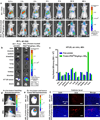
(a–c) Systemic delivery. (a,b) In vivo (a) and ex vivo (b) animal images of the HT-29 colon cancer bearing nude mice xenograft models (#A1 and #A2) after tail vein injection of free Cy5-insulin, and Cy5-insulin-loaded PEG5k(ArgArg-L-VE)4 NPs at a loading ratio of 1/5 (P/T, w/w). The black circles in (a) indicate the tumor sites. (c) Quantitative analysis of the ex vivo tumor and organ uptake. (d–f) Local delivery. (d) In vivo bioluminescence images for brain tumor tracking in the mice (#B1 and #B2) injected with intracranial U87 tumors taken using the Photon Imager system at day 7 post injection. (e,f) Distributions in intracranial U87 tumors determined by ex vivo imaging (e) and at the cellular level determined by microscopy (f) at 48 h after CED of free pHLIP-Alexa750-DOX, and pHLIP-Alexa750-DOX-loaded telodendrimer NPs at a loading ratio of 1/5 (peptide/telodendrimer, w/w). The cell nuclei in (f) were stained with DAPI (blue).

Similar articles

Cited by

References

    1. Leader B, Baca QJ, Golan DE. Protein therapeutics: a summary and pharmacological classification. Nat. Rev. Drug. Discov. 2008;7:21–39. - PubMed
    1. Wang C-Y, Mayo MW, Baldwin AS. TNF-and cancer therapy-induced apoptosis: potentiation by inhibition of NF-κB. Science. 1996;274:784–787. - PubMed
    1. Chen J, Zhao M, Feng F, Sizovs A, Wang J. Tunable thioesters as “reduction” responsive functionality for traceless reversible protein PEGylation. J. Am. Chem. Soc. 2013;135:10938–10941. - PubMed
    1. Liu M, Johansen P, Zabel F, Leroux J-C, Gauthier MA. Semi-permeable coatings fabricated from comb-polymers efficiently protect proteins in vivo. Nat. Commun. 2014;5:5526. - PubMed
    1. Kobsa S, Saltzman WM. Bioengineering approaches to controlled protein delivery. Pediatr. Res. 2008;63:513–519. - PubMed

Publication types

LinkOut - more resources