. 2016 Jul;22(7):807-11.
doi: 10.1038/nm.4124. Epub 2016 Jun 13.

Stimulating the RIG-I pathway to kill cells in the latent HIV reservoir following viral reactivation

Affiliations

Stimulating the RIG-I pathway to kill cells in the latent HIV reservoir following viral reactivation

Peilin Li et al. Nat Med. 2016 Jul.

Abstract

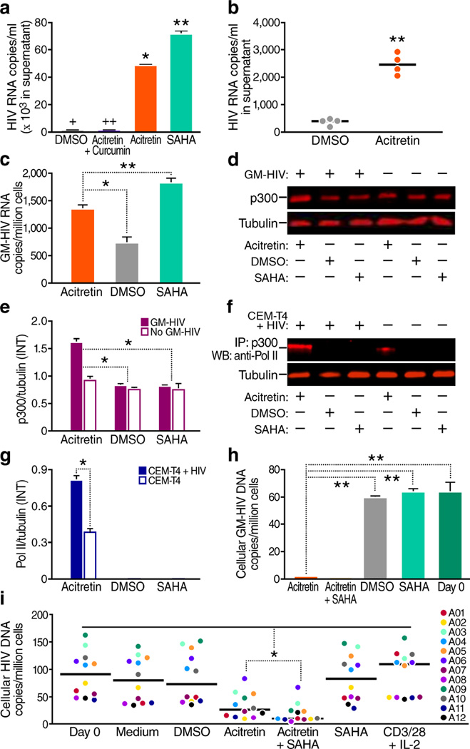
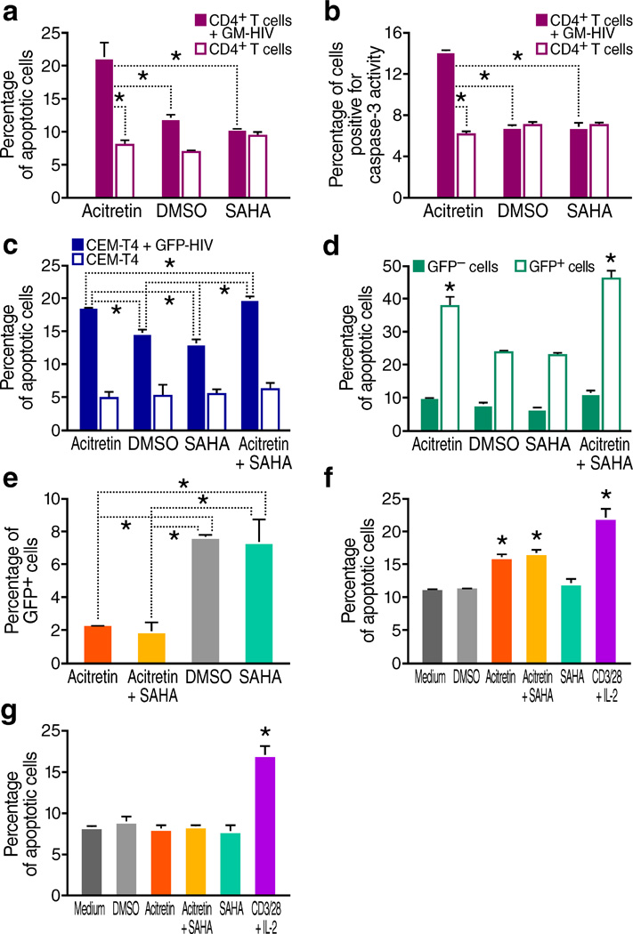
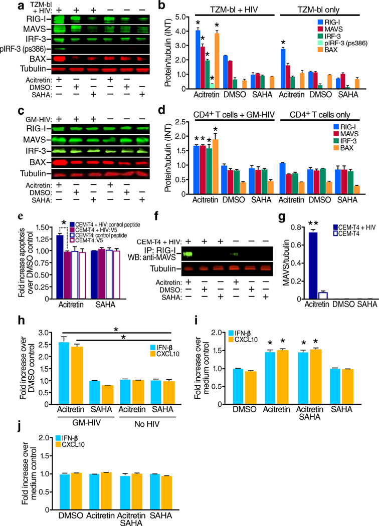
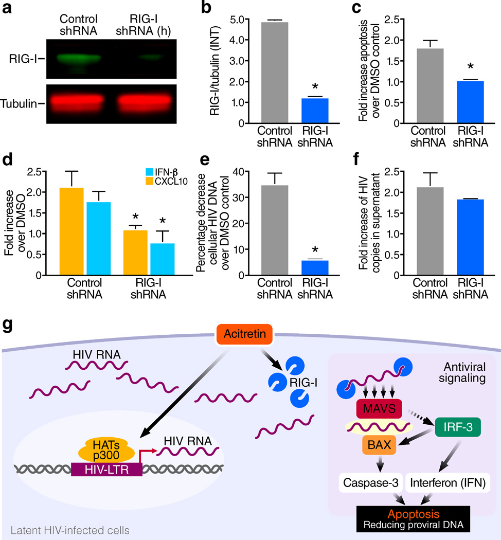
The persistence of latent HIV proviruses in long-lived CD4(+) T cells despite antiretroviral therapy (ART) is a major obstacle to viral eradication. Because current candidate latency-reversing agents (LRAs) induce HIV transcription, but fail to clear these cellular reservoirs, new approaches for killing these reactivated latent HIV reservoir cells are urgently needed. HIV latency depends upon the transcriptional quiescence of the integrated provirus and the circumvention of immune defense mechanisms. These defenses include cell-intrinsic innate responses that use pattern-recognition receptors (PRRs) to detect viral pathogens, and that subsequently induce apoptosis of the infected cell. Retinoic acid (RA)-inducible gene I (RIG-I, encoded by DDX58) forms one class of PRRs that mediates apoptosis and the elimination of infected cells after recognition of viral RNA. Here we show that acitretin, an RA derivative approved by the US Food and Drug Administration (FDA), enhances RIG-I signaling ex vivo, increases HIV transcription, and induces preferential apoptosis of HIV-infected cells. These effects are abrogated by DDX58 knockdown. Acitretin also decreases proviral DNA levels in CD4(+) T cells from HIV-positive subjects on suppressive ART, an effect that is amplified when combined with suberoylanilide hydroxamic acid (SAHA), a histone deacetylase inhibitor. Pharmacological enhancement of an innate cellular-defense network could provide a means by which to eliminate reactivated cells in the latent HIV reservoir.


Conflict of interest statement

The authors declare that they have no conflict of interest.

Figures

Figure 1
Figure 1
Acitretin induces HIV expression and reduces cellular HIV-DNA in CD4+ T cells. (a) HIV copy number in the supernatant of a latent HIV T-cell line (ACH-2) after 72 h of treatment. Both acitretin (P < 0.05) and SAHA significantly increased HIV transcription (*P < 0.01), and curcumin (an inhibitor of p300) limited acitretin induction of HIV. (+HIV ‘copy number, 486.6 ± 5.9’), (++HIV ‘copy number, 379.6 ± 17.8’). (b) HIV-RNA copy number in supernatants from cultures of CD4+ T cells from four aviremic HIV+ subjects on ART at day 6. Acitretin significantly increased HIV transcription (P < 0.01 versus. DMSO control). (c) Cellular GM-HIV-RNA copies/million cells after 24 h of the indicated treatment of infected primary CD4+ T cells. Both acitretin and SAHA increased GM-HIV transcription to a greater extent than DMSO. The increase was greater with SAHA than acitretin (P < 0.01). (d) Immunoblot analysis of p300 and tubulin proteins from both GM-HIV-infected and uninfected CD4+ T cells (from the same donor) after 48 h of treatment. (e) The ratio of mean value intensities (INT) for p300 and tubulin from panel (d, n = 4) confirming significantly higher expression of p300 in infected cells treated with acitretin than in cells treated with DMSO or SAHA. (f) Immunoblot analysis of co-immunoprecipitation of protein extracts of CEM-T4 cells with or without latent GFP-HIV virus using antibody against p300 and western blot for RNA pol II after 48 h of treatment. Association of p300 with RNA pol II is enhanced by acitretin. (g) The ratio of RNA pol II to tubulin from (f, n = 4) is greatest with acitretin treatment of cells with GFP-HIV. (h) GM-HIV-DNA content in cellular DNA after 72 h of treatment. GM-HIV-DNA was significantly lower after treatment with acitretin or acitretin plus SAHA than with SAHA or DMSO (P < 0.001). GM-HIV-DNA was not detectable despite testing of cellular DNA from two million cells after treatment with acitretin plus SAHA. (i) HIV-DNA concentrations at day 7 of treatment in CD4+ T cells from HIV+ subjects on ART (n = 12). Both acitretin and acitretin plus SAHA significantly lowered HIV-DNA concentrations in cells from all 12 HIV+ subjects (P < 0.05 compared to treatment with DMSO, SAHA, medium, or anti-CD3 and anti-CD28 antibodies beads plus IL-2 (CD3/28+IL-2). HIV-DNA concentrations were significantly lower after treatment with acitretin plus SAHA than after treatment with acitretin alone (P < 0.05). Values represent mean ± s.e.m. of duplicate samples from HIV+ subjects (b,i), and triplicate samples from the ACH-2 (a) and GM-HIV infection model(c, h) from three independent experiments. A student's t-Test was used to compare experimental conditions (a, b, c, e, g, h, i); *P<0.05; **P<0.01.
Figure 2
Figure 2
Acitretin preferentially induces apoptosis in HIV-infected cells. (a) Percentage of apoptotic cells determined by annexin V staining of GM-HIV-infected or mock-infected CD4+Tcells after 72 h of treatment. The percentage of apoptotic cells was significantly greater in the presence of acitretin than with SAHA or DMSO in GM-HIV-infected but not uninfected CD4+T cells (P < 0.05). (b) Percentage of cells expressing active caspase-3 determined by flow cytometry assessed 72 h of treatment with DMSO, SAHA or acitretin. Caspase-3 activity was preferentially increased in GM-HIV infected CD4+T cells treated with acitretin (P<0.05). (c) Percentage of apoptotic cells determined by annexin V staining of GFP-HIV-infected and uninfected CEM-T4 cells. Acitretin and acitretin plus SAHA increased apoptosis at 48 h in GFP-HIV-infected cells to a significantly greater extent than SAHA or DMSO alone (P <0.05) although both of these agents produced higher than expected levels of cell death in infected cells compared to uninfected cells. No differences were detected in uninfected CEM-T4 cells. (d) Percentage of apoptotic cells determined by annexin V staining of infected cells from (c) gated for the presence or absence of expression of GFP encoded by the reporter virus. Acitretin and acitretin plus SAHA increased apoptosis of GFP-positive cells to a significantly greater degree than SAHA or DMSO, while no significant increases were induced by these agents in GFP negative cells (P < 0.05). (e) Percentage of GFP-positive cells after 7 d of treatment, as determined by flow cytometry of infected cells from (c). Both acitretin and acitretin plus SAHA reduced the number of GFP-HIV-positive cells significantly more (P < 0.05) than treatment with SAHA or DMSO. (f) Average percentage of apoptotic cells determined by annexin V staining of CD4+ T cells from HIV+ subjects on ART (n = 12). After 7 d of treatment with acitretin and acitretin plus SAHA, the frequency of apoptotic cells was significantly increased in patient CD4+ T cells compared to DMSO, SAHA, or medium (P < 0.05). Apoptosis was also significantly increased following treatment with anti-CD3 and anti-CD28 antibodies beads plus IL-2 (CD3/28+IL-2) (P < 0.05). (g) Mean percentage of apoptotic cells determined by annexin V staining in cultures of CD4+ T cells from four healthy control subjects. Neither acitretin, nor acitretin plus SAHA induced higher levels of apoptosis than DMSO, SAHA, or medium at day 7 in these normal cells while increased apoptosis was observed when these normal cells were treated with anti-CD3 and anti-CD28 antibodies beads plus IL-2(CD3/28+IL-2) (P < 0.05). Values represent mean ± s.e.m. of duplicate samples from experiments with cells from 12 HIV+ subjects (f), values represent mean ± s.e.m. of triplicate samples for (a,b,c,d,e) from three independent experiments, and values represent mean ± s.e.m.of triplicate samples for (g) from four healthy donor CD4 T cells experiments. A student's t-Test was used to compare experimental conditions (a–g); **P < 0.01, *P < 0.05.
Figure 3
Figure 3
Acitretin increases expression of RIG-I signaling pathway proteins including MAVS, IRF3, p-IRF3, and BAX, and increases production of IFN-β, and CXCL10 in cells infected with HIV. (a) Representative immunoblot analysis of RIG-I, MAVS, IRF3, p-IRF3 (antibody detecting phosphorylation at Ser 386), BAX, and tubulin at 48 h following treatment of HIV infected or uninfected TZM-bl cells with acitretin, DMSO or SAHA. (b) The ratio of mean value intensities (INT) for each protein and tubulin for the immunoblot bands in (a) combined with results from three additional independent experiments (n = 4). (c) Representative immunoblot analysis of RIG-I, MAVS, IRF3, BAX. and tubulin at 48 h after acitretin treatment of GM- HIV-infected and uninfected CD4+ T cells with acitretin, DMSO or SAHA. (d) The ratio of mean value intensities (INT) of each protein and tubulin for the immunoblot bands in panel (c) combined with results from three additional independent experiments (n = 4). RIG-I, MAVS, IRF3,p-IRF3 and BAX significant increased treated with acitretin compare to SAHA or MDSO control (P<0.05). RIG-I was also increased by acitretin in uninfected TZM-bl cells and reached significant (P<0.05)(b), but in uninfected CD4+ T cells from the same donors this increase did not reach statistical significance when compared to the DMSO control(d). (e) Fold increases in annexin V positive, apoptotic cells in uninfected or HIV-infected CEM-T4 cells treated with acitretin or SAHA (versus DMSO) in the presence of the V5 peptide (a BAX inhibitor) or a control peptide. Note significant decreases in apoptosis production by the V5 peptide (a BAX inhibitor) in acitretin treated infected cells (P<0.05) but not in the other conditions. (f, g) Immunoblot analysis of co-immunoprecipitation of protein extracts of CEM-T4 cells with or without latent GFP-HIV virus using antibody against RIG-I followed by immunoblotting for MAVS after 48 h of treatment with acitretin, DMSO, or SAHA. Note increased association of RIG-I with MAVS in infected cells treated with acitretin. (g) The association of MAVS with RIG-I following acitretin treatment in (f, n = 4) was higher in GFP-HIV infected cells than in uninfected cells. (h) Fold increase over DMSO control in type 1 interferon (IFN-β) and CXCL10 expression in supernatants from GM-HIV-infected, and uninfected CD4+ T cells 72 h after treatment. Acitretin increased IFN-β and CXCL10 levels to a greater extent than SAHA (P < 0.05) in infected but not uninfected cells. (i) Fold increase over medium control of IRF3-induced type 1 interferon (IFN-β) and CXCL10 expression in supernatants of cultures of CD4+ T cells from HIV+ subjects on ART (n = 12) on day 7 of treatment. Both acitretin and acitretin plus SAHA significantly induced IFN-β and CXCL10 (P < 0.05 vs. SAHA and DMSO). (j) No differences in IFN-β and CXCL10 expression occurred in healthy control CD4+ T cells treated with DMSO, acitretin, SAHA or acitretin plus SAHA. All values represent mean ± s.e.m. of duplicate samples from HIV+ subjects(i), and values represent mean ± s.e.m. of triplicate samples for (e,h) from three independent experiments, and values represent mean ± s.e.m.of triplicate samples for (j) from four healthy donor CD4 T cells experiments. A student's t-Test was used to compare experimental conditions (b, d, e, g, h, i, j); **P < 0.01, *P < 0.05.
Figure 4
Figure 4
Short hairpin RNA (shRNA) knockdown of RIG-I expression markedly inhibits acitretin enhancement of apoptosis, induction of IFN-β and CXCL10 production and preferential depletion of HIV-DNA+ cells. CEM T4 cells were transfected with either RIG-I-shRNA plasmid DNA, or control-shRNA (scrambled shRNA) plasmid DNA, cultured for 72 h and then selected in the presence of puromycin (10µg/ml) for 7 d. Next, these cells were infected with HIV-NL43 and maintained for 7 d with antiviral drugs as previously described for the in vitro latency model. (a) Immunoblot analysis of RIG-I and tubulin after the puromycin selection, (b) the ratio of mean value intensities (INT) of RIG-I to tubulin from (a, n = 4), RIG-I shRNA transfection significantly reduced RIG-I expression (P<0.05).(c) Knockdown of RIG-I significantly diminished (P<0.05) acitretin induced apoptosis at day 7 post treatment compared to the scrambled shRNA control, and (d) significantly reduced (P<0.05) acitretin induced CXCL10 and IFN-β release and (e) significantly impaired acitretin-induced decreases in HIV-DNA levels (P<0.05). (f) Knockdown of RIG-I expression conversely had no significant impact on acitretin-stimulated release of HIV-RNA into supernatant at day 7, the fold increase of HIV copies in supernatant are from acitretin compared to DMSO control in both conditions. All values represent mean ± s.e.m. of triplicate samples (b,c,d,e,f) from three independent experiments. A student's t-Test was used to compare experimental conditions (b–f); **P < 0.01, *P < 0.05. (g) Model depicting the preferential enhancement of RIG-I signaling by acitretin in latently infected HIV cells. Acitretin increases both RIG-I and HIV-RNA expression in the cytoplasm of latent HIV-infected cells; recognition of HIV-RNA by RIG-I initiates antiviral signaling. RIG-I–RIG-I interactions produce conformational changes that permit binding to MAVS, which leads in turn promotes IRF-3 activation. Activated IRF-3 activates two pathways: one inducing type I interferon (IFN) production and another resulting in the binding of BAX and activation of caspase-3-dependent apoptosis in the virally infected cell. These two pathways work together to protect the host against further spread of the pathogen. LTR, long terminal repeat.

References

    1. Wong JK, et al. Recovery of replication-competent HIV despite prolonged suppression of plasma viremia. Science. 1997;278:1291–1295. - PubMed
    1. Finzi D, et al. Identification of a reservoir for HIV-1 in patients on highly active antiretroviral therapy. Science. 1997;278:1295–1300. - PubMed
    1. Chun TW, et al. Presence of an inducible HIV-1 latent reservoir during highly active antiretroviral therapy. Proc Natl Acad Sci U S A. 1997;94:13193–13197. - PMC - PubMed
    1. Deeks SG, Lewin SR, Havlir DV. The end of AIDS: HIV infection as a chronic disease. Lancet. 2013;382:1525–1533. - PMC - PubMed
    1. Passaes CP, Saez-Cirion A. HIV cure research: advances and prospects. Virology. 2014;454–455:340–352. - PubMed

MeSH terms