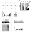
ARTD1 regulates cyclin E expression and consequently cell-cycle re-entry and G1/S progression in T24 bladder carcinoma cells
- PMID: 27295004
- PMCID: PMC4968958
- DOI: 10.1080/15384101.2016.1195530
ARTD1 regulates cyclin E expression and consequently cell-cycle re-entry and G1/S progression in T24 bladder carcinoma cells
Abstract
ADP-ribosylation is involved in a variety of biological processes, many of which are chromatin-dependent and linked to important functions during the cell cycle. However, any study on ADP-ribosylation and the cell cycle faces the problem that synchronization with chemical agents or by serum starvation and subsequent growth factor addition already activates ADP-ribosylation by itself. Here, we investigated the functional contribution of ARTD1 in cell cycle re-entry and G1/S cell cycle progression using T24 urinary bladder carcinoma cells, which synchronously re-enter the cell cycle after splitting without any additional stimuli. In synchronized cells, ARTD1 knockdown, but not inhibition of its enzymatic activity, caused specific down-regulation of cyclin E during cell cycle re-entry and G1/S progression through alterations of the chromatin composition and histone acetylation, but not of other E2F-1 target genes. Although Cdk2 formed a functional complex with the residual cyclin E, p27(Kip 1) protein levels increased in G1 upon ARTD1 knockdown most likely due to inappropriate cyclin E-Cdk2-induced phosphorylation-dependent degradation, leading to decelerated G1/S progression. These results provide evidence that ARTD1 regulates cell cycle re-entry and G1/S progression via cyclin E expression and p27(Kip 1) stability independently of its enzymatic activity, uncovering a novel cell cycle regulatory mechanism.
Keywords: E2F-1; PARP1; cell cycle regulation; cyclin E; gene expression; p27.
Figures


Similar articles
-
PARP1 promoter links cell cycle progression with adaptation to oxidative environment.Redox Biol. 2018 Sep;18:1-5. doi: 10.1016/j.redox.2018.05.017. Epub 2018 Jun 2. Redox Biol. 2018. PMID: 29886395 Free PMC article. Review.
-
Depletion of SUMO ligase hMMS21 impairs G1 to S transition in MCF-7 breast cancer cells.Biochim Biophys Acta. 2012 Dec;1820(12):1893-900. doi: 10.1016/j.bbagen.2012.08.002. Epub 2012 Aug 10. Biochim Biophys Acta. 2012. PMID: 22906975
-
Knockdown of protein tyrosine phosphatase SHP-1 inhibits G1/S progression in prostate cancer cells through the regulation of components of the cell-cycle machinery.Oncogene. 2010 Jan 21;29(3):345-55. doi: 10.1038/onc.2009.329. Epub 2009 Oct 19. Oncogene. 2010. PMID: 19838216
-
Phospholipase C delta 1 regulates cell proliferation and cell-cycle progression from G1- to S-phase by control of cyclin E-CDK2 activity.Biochem J. 2008 Nov 1;415(3):439-48. doi: 10.1042/BJ20080233. Biochem J. 2008. PMID: 18588506
-
Low-Molecular-Weight Cyclin E in Human Cancer: Cellular Consequences and Opportunities for Targeted Therapies.Cancer Res. 2018 Oct 1;78(19):5481-5491. doi: 10.1158/0008-5472.CAN-18-1235. Epub 2018 Sep 7. Cancer Res. 2018. PMID: 30194068 Free PMC article. Review.
Cited by
-
PARP1 promoter links cell cycle progression with adaptation to oxidative environment.Redox Biol. 2018 Sep;18:1-5. doi: 10.1016/j.redox.2018.05.017. Epub 2018 Jun 2. Redox Biol. 2018. PMID: 29886395 Free PMC article. Review.
-
Dysregulated proliferation and immune response induced by estrogen in Egr1 knockout uterus are similar to those in immature uterus.BMC Genomics. 2025 Aug 1;26(1):715. doi: 10.1186/s12864-025-11904-3. BMC Genomics. 2025. PMID: 40751132 Free PMC article.
-
Cell Synchronization by Double Thymidine Block.Bio Protoc. 2018 Sep 5;8(17):e2994. doi: 10.21769/BioProtoc.2994. Bio Protoc. 2018. PMID: 30263905 Free PMC article.
-
Effects of three IL-15 variants on NCI-H446 cell proliferation and expression of cell cycle regulatory molecules.Oncotarget. 2017 Nov 20;8(64):108108-108117. doi: 10.18632/oncotarget.22550. eCollection 2017 Dec 8. Oncotarget. 2017. PMID: 29296227 Free PMC article.
-
Parvoviruses NS1 oncolytic attributes: mechanistic insights and synergistic anti-tumor therapeutic strategies.Front Microbiol. 2025 Aug 11;16:1631433. doi: 10.3389/fmicb.2025.1631433. eCollection 2025. Front Microbiol. 2025. PMID: 40862157 Free PMC article. Review.
References
-
- Murray AH, Hunt T. The cell cycle: an introduction. New York: Oxford University Press, 1993.
-
- Ren S, Rollins BJ. Cyclin C/cdk3 promotes Rb-dependent G0 exit. Cell 2004; 117:239-51; PMID:15084261; http://dx.doi.org/10.1016/S0092-8674(04)00300-9 - DOI - PubMed
-
- Rissland OS, Hong SJ, Bartel DP. MicroRNA destabilization enables dynamic regulation of the miR-16 family in response to cell-cycle changes. Mol Cell 2011; 43:993-1004; PMID:21925387; http://dx.doi.org/10.1016/j.molcel.2011.08.021 - DOI - PMC - PubMed
-
- Sage J. Cyclin C makes an entry into the cell cycle. Dev Cell 2004; 6:607-8; PMID:15130482; http://dx.doi.org/10.1016/S1534-5807(04)00137-6 - DOI - PubMed
MeSH terms
Substances
LinkOut - more resources
Full Text Sources
Other Literature Sources
Medical
Miscellaneous
