. 2017 Aug;56(5):1919-1930.
doi: 10.1007/s00394-016-1234-9. Epub 2016 Jun 13.

Characterizing microbiota-independent effects of oligosaccharides on intestinal epithelial cells: insight into the role of structure and size : Structure-activity relationships of non-digestible oligosaccharides

Affiliations

Characterizing microbiota-independent effects of oligosaccharides on intestinal epithelial cells: insight into the role of structure and size : Structure-activity relationships of non-digestible oligosaccharides

Peyman Akbari et al. Eur J Nutr. 2017 Aug.

Abstract

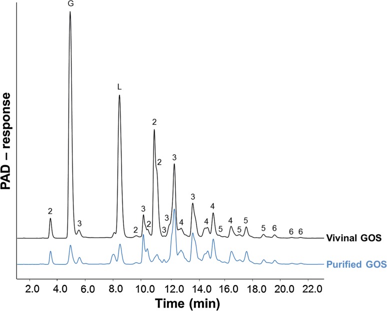
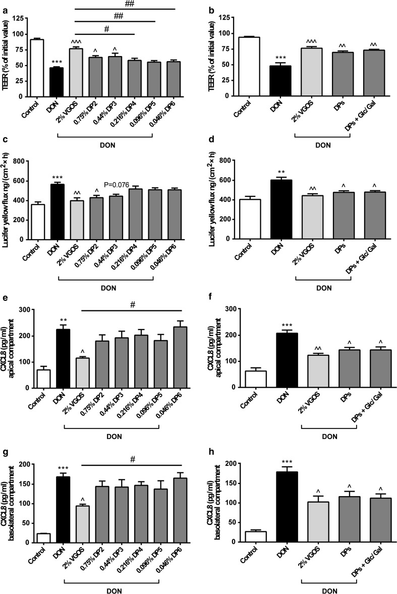
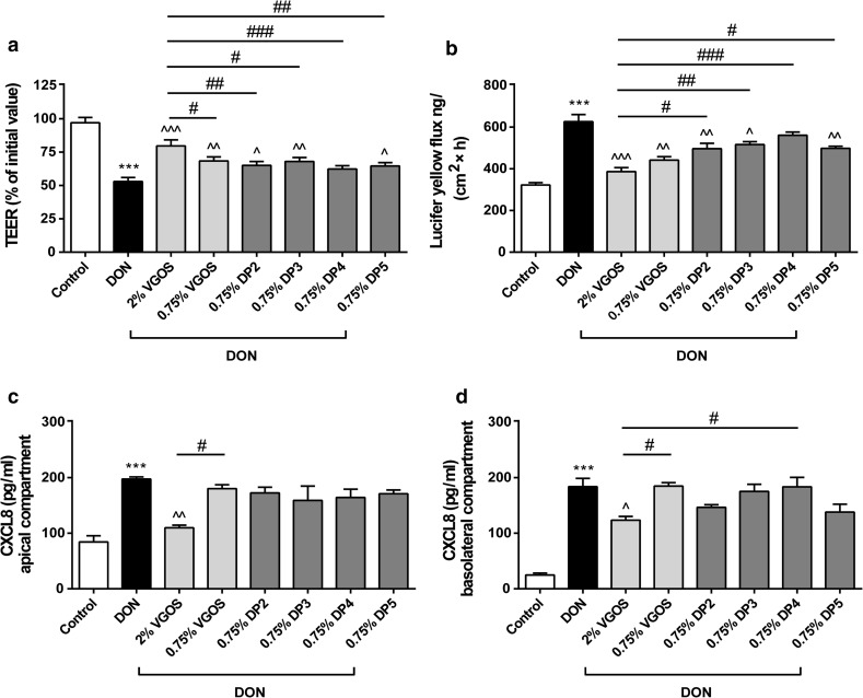
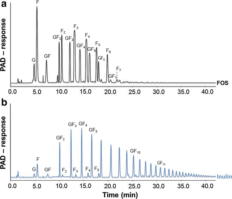
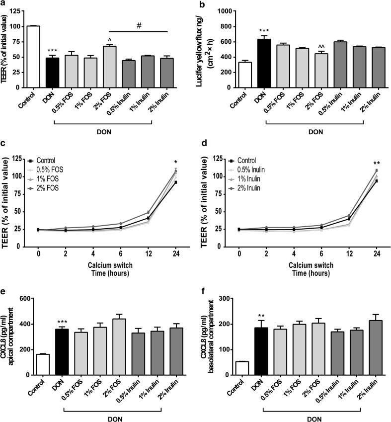
Purpose: The direct effects of galacto-oligosaccharides (GOS), including Vivinal® GOS syrup (VGOS) and purified Vivinal® GOS (PGOS), on the epithelial integrity and corresponding interleukin-8 (IL-8/CXCL8) release were examined in a Caco-2 cell model for intestinal barrier dysfunction. To investigate structure-activity relationships, the effects of individual DP fractions of VGOS were evaluated. Moreover, the obtained results with GOS were compared with Caco-2 monolayers incubated with fructo-oligosaccharides (FOS) and inulin.

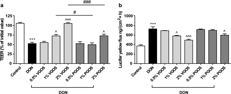
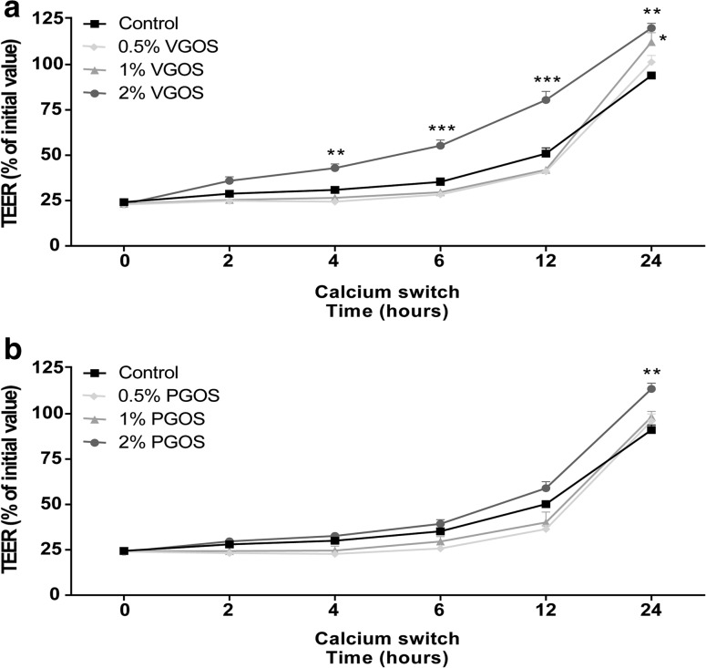
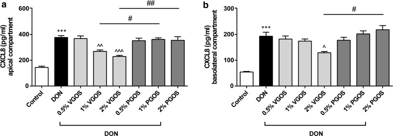
Methods: Caco-2 monolayers were pretreated (24 h) with or without specific oligosaccharides or DP fractions of VGOS (DP2 to DP6) before being exposed for 12 or 24 h to the fungal toxin deoxynivalenol (DON). Transepithelial electrical resistance and lucifer yellow permeability were measured to investigate barrier integrity. A calcium switch assay was used to study the reassembly of tight junction proteins. Release of CXCL8, a typical marker for inflammation, was quantified by ELISA.

Results: In comparison with PGOS, FOS and inulin, VGOS showed the most pronounced protective effect on the DON-induced impairment of the monolayer integrity, acceleration of the tight junction reassembly and the subsequent CXCL8 release. DP2 and DP3 in concentrations occurring in VGOS prevented the DON-induced epithelial barrier disruption, which could be related to their high prevalence in VGOS. However, no effects of the separate DP GOS fractions were observed on CXCL8 release.

Conclusions: This comparative study demonstrates the direct, microbiota-independent effects of oligosaccharides on the intestinal barrier function and shows the differences between individual galacto- and fructo-oligosaccharides. This microbiota-independent effect of oligosaccharides depends on the oligosaccharide structure, DP length and concentration.

Keywords: CXCL8; Caco-2 cells; Degree of polymerization; Intestinal permeability; Non-digestible oligosaccharides; Tight junctions.


Conflict of interest statement

Akbari, Schols and Fink-Gremmels have no interest to declare. Willems, Difilippo and Braber were granted by the Carbohydrate Competence Center (CCC) project as indicated in the funding sources; Garssen is associated with Nutricia Research and Schoterman with FrieslandCampina, respectively, both of which are industrial partners in the Dutch Carbohydrate Competence Center project.

Figures

Fig. 1
Fig. 1
VGOS and PGOS characteristics. HPAEC-PAD elution patterns of Vivinal® GOS syrup and purified GOS. Numbers 26 correspond to galacto-oligosaccharides having a degree of polymerization from 2 to 6. G and L represent galactose/glucose and lactose, respectively
Fig. 2
Fig. 2
Different effects of VGOS and PGOS on the DON-induced impairment of the Caco-2 cell monolayer integrity. Caco-2 cells were pretreated apically and basolaterally with increasing concentrations (0.5, 1 and 2 %) of VGOS or PGOS (24 h) prior to the addition of DON (4.2 μM) (apical and basolateral compartments) for another 24 h. Subsequently, the TEER (a) and the translocation of lucifer yellow from the apical to the basolateral compartment (b) were measured. Results are expressed as a percentage of initial value (TEER) or the amount of tracer transported [ng/(cm2 × h)] as mean ± SEM of three independent experiments, each performed in triplicate (***P < 0.001: significantly different from the unstimulated cells; ^P < 0.05, ^^^P < 0.001: significantly different from the DON-stimulated cells; # P < 0.05, ### P < 0.001: significantly different from each other)
Fig. 3
Fig. 3
VGOS time-dependently accelerated tight junction reassembly after calcium deprivation in Caco-2 cells. Caco-2 cells were pretreated apically and basolaterally with increasing concentrations (0.5, 1 and 2 %) of VGOS (a) or PGOS (b) (24 h) prior to transient calcium deprivation with HBSS-EGTA to disrupt tight junction proteins. TEER was measured at the indicated time points (0, 2, 4, 6, 12 and 24 h) during recovery in complete, calcium-containing DMEM with either VGOS (a) or PGOS (b). Results are expressed as a percentage of initial value as mean ± SEM of three independent experiments, each performed in triplicate (*P < 0.05, **P < 0.01, ***P < 0.001: significantly different from the untreated cells)
Fig. 4
Fig. 4
VGOS was able to suppress the DON-induced increase in CXCL8 release by Caco-2 cells. Caco-2 cells were pretreated apically and basolaterally with increasing concentrations (0.5, 1 and 2 %) of VGOS or PGOS (24 h) prior to the addition of DON (4.2 μM) (apical and basolateral compartments) for 24 h. CXCL8 secretion into medium of apical (a) and basolateral (b) compartments was measured by ELISA. Results are expressed as pg/ml as mean ± SEM of three independent experiments, each performed in triplicate (***P < 0.001; significantly different from the unstimulated cells. ^P < 0.05, ^^P < 0.01, ^^^P < 0.001: significantly different from the DON-stimulated cells; # P < 0.05, ## P < 0.01: significantly different from each other)
Fig. 5
Fig. 5
Combined DP fractions of VGOS mimicked VGOS in preventing DON-induced barrier disruption and CXCL8 release, whereas only individual DP2 and DP3 can prevent DON-induced barrier disruption. Caco-2 cells were pretreated apically and basolaterally with VGOS, individual DP fractions of GOS (ranging from DP2 to DP6) and combination of different DP fractions (DP2–DP6) with or without supplementation with glucose (Glc) and galactose (Gal) (24 h) prior to the addition of DON (4.2 μM) (apical and basolateral compartments) for 12 h. Subsequently, TEER (a, b), the transport of lucifer yellow (c, d) and CXCL8 release into the apical (e, f) and basolateral (g, h) compartment were measured. Results are expressed as a percentage of initial value (TEER), the amount of tracer transported [ng/(cm2 × h)] or pg/ml CXCL8 as mean ± SEM of three independent experiments, each performed in triplicate (**P < 0.01, ***P < 0.001: significantly different from the unstimulated cells; ^P < 0.05, ^^P < 0.01, ^^^P < 0.001: significantly different from the DON-stimulated cells; # P < 0.05, ## P < 0.01: significantly different from each other)
Fig. 6
Fig. 6
Different effects on the Caco-2 cell monolayer induced by individual DP fractions of VGOS with equal concentrations. Caco-2 cells were pretreated apically and basolaterally with VGOS (0.75 and 2 %) or individual DP fractions of VGOS (0.75 %, ranging from DP2 to DP5) (24 h) prior to the addition of DON (4.2 μM) (apical and basolateral compartments) for 12 h. Subsequently, TEER (a), the transport of lucifer yellow (b) and CXCL8 secretion into medium of apical (c) and basolateral (d) compartments were measured. Results are expressed as a percentage of initial value (TEER), the amount of tracer transported [ng/(cm2 × h)] or pg/ml CXCL8 as mean ± SEM and are representative of two independent experiments, each performed in triplicate (***P < 0.001: significantly different from the unstimulated cells; ^P < 0.05, ^^P < 0.01, ^^^P < 0.001: significantly different from the DON-stimulated cells; # P < 0.05, ## P < 0.01, ### P < 0.001: significantly different from each other)
Fig. 7
Fig. 7
FOS and inulin characteristics. HPAEC-PAD elution patterns of FOS (a) and inulin (b). The F n series represent oligomers consisting of fructose only, whereas the GF n series represent fructose oligomers terminated with a terminal glucose molecule. The number of fructose units within an oligomer is indicated by n
Fig. 8
Fig. 8
Different effects of FOS and inulin on the DON-induced barrier disruption, tight junction reassembly and CXCL8 release. Caco-2 cells were pretreated apically and basolaterally with increasing concentrations (0.5, 1 and 2 %) of FOS or inulin (24 h) prior to the addition of DON (4.2 μM) (apical and basolateral compartments) for another 24 h (a, b, e, f) or transient calcium deprivation with HBSS-EGTA to disrupt tight junction proteins (c, d). Subsequently, TEER values at the indicated time points (a, c, d), the transport of lucifer yellow (b) and CXCL8 secretion into medium of apical (e) and basolateral (f) compartments were measured. Results are expressed as a percentage of initial value (TEER), the amount of tracer transported [ng/(cm2 × h)] or pg/ml CXCL8 as mean ± SEM of three independent experiments, each performed in triplicate (*P < 0.05, **P < 0.01, ***P < 0.001: significantly different from the unstimulated cells; ^P < 0.05, ^^P < 0.01: significantly different from the DON-stimulated cells; # P < 0.05: significantly different from each other)

References

    1. Bode L. The functional biology of human milk oligosaccharides. Early Hum Dev. 2015;91:619–622. doi: 10.1016/j.earlhumdev.2015.09.001. - DOI - PubMed
    1. Boehm G, Stahl B. Oligosaccharides from milk. J Nutr. 2007;137:S847–S849. - PubMed
    1. Boehm G, Fanaro S, Jelinek J, Stahl B, Marini A. Prebiotic concept for infant nutrition. Acta Paediatr. 2003;91:64–67. - PubMed
    1. Fanaro S, Boehm G, Garssen J, Knol J, Mosca F, Stahl B, Vigi V. Galacto-oligosaccharides and long-chain fructo-oligosaccharides as prebiotics in infant formulas: a review. Acta Paediatr. 2005;94:22–26. doi: 10.1080/08035320510043538. - DOI - PubMed
    1. Ben XM, Zhou XY, Zhao WH, Yu WL, Pan W, Zhang WL, Wu SM, Van Beusekom CM, Schaafsma A. Supplementation of milk formula with galacto-oligosaccharides improves intestinal micro-flora and fermentation in term infants. Chin Med J (Engl) 2004;117:927–931. - PubMed